
Sustainable Manufacturing and Foundry Practices


1Department of Production and Industrial Engineering, National Institute of Technology Jamshedpur, Jharkhand, India

This article is distributed under the terms of the Creative Commons Attribution-NonCommercial 4.0 License (https://creativecommons.org/licenses/by-nc/4.0/) which permits non-commercial use, reproduction and distribution of the work without further permission provided the original work is attributed as specified on the SAGE and Open Access page (https://us.sagepub.com/en-us/nam/open-access-at-sage).
Magnesium-based biodegradable composites offer significant advantages over nondegradable materials for biomedical implants. These materials degrade gradually in physiological environments, thereby reducing stress shielding and eliminating the need for secondary implant removal surgeries. This review explores how alloying elements (e.g., Zn, Ca, Sr) in the magnesium matrix and reinforcing phases (e.g., hydroxyapatite, SiC, TiC) in metal matrix composites influence degradation kinetics, mechanical integrity, and ion release behaviour. Proper regulation of the degradation rate is critical for maintaining mechanical support during the healing phase while ensuring biocompatibility. This study expands the use of magnesium composites in implantable medical devices. Some examples are orthopaedic implants, circulatory stents, bone fixation devices, and soft tissue scaffolds. The above uses demonstrate the ability of magnesium composites to improve patient outcomes and reduce implant-related procedures through gradual breakdown and tissue regeneration. The review also reports the biodegradation control, biocompatibility, manufacturing scale-up, and regulatory requirements for clinical translation. Successful commercialisation depends on balancing the degradation rate with mechanical support, limiting cytotoxicity, and meeting regulatory standards. In conclusion, magnesium-based biodegradable composites have the potential to revolutionise biomedical implants. The unique combination of mechanical properties, biocompatibility, and regulated biodegradation improves patient outcomes and therapy efficacy. Medical device design and regenerative medicine will benefit from magnesium composites as researchers improve synthesis methods, material properties, and overcome barriers.
Biodegradable, alloying elements, reinforcement, osteogenesis, corrosion, cytotoxicity
Introduction
Biodegradable magnesium-based metal matrix composites (Mg-MMCs) represent a paradigm shift in temporary implant design for bone fractures, bone defects, and cardiovascular applications, addressing key limitations of traditional permanent implants such as stress shielding, chronic inflammation, and the need for revision surgeries (Chandra & Pandey, 2020; Kim et al., 2020; Tsakiris et al., 2021; Zhang et al., 2021). Unlike nondegradable metals (e.g., titanium and stainless steel), Mg-MMCs degrade progressively in physiological environments, ideally matching the rate of tissue healing while providing temporary mechanical support (Choudhary & Singh Raman, 2015; Fu et al., 2011; Seetharaman et al., 2023; Sheikh et al., 2015). This review critically synthesises recent advances in synthesis, reinforcement strategies, degradation control, and clinical translation of Mg-MMCs (Bairagi & Mandal, 2022; Bommala et al., 2019; Haghshenas, 2017; Zhao et al., 2017).
Temporary implants are widely used in bone fixation, tissue scaffolding, and stent implantation. Orthopaedic implants play a critical role in treating bone fractures and defects by providing temporary support and stability during the healing process. The bone plate is the most common internal fixation device, secured to bone fragments with screws (Kim et al., 2020). Magnesium alloys and Mg-MMCs are highly attractive for biomedical applications due to their low density, high specific strength, and excellent biocompatibility, making them suitable for use in aerospace, automotive, and medical fields (Bairagi & Mandal, 2022; Choudhary & Singh Raman, 2015; Zhao et al., 2017).
The capacity of biodegradable materials to undergo slow degradation and subsequent resorption by the human body is in accordance with the inherent tissue repair mechanism, hence facilitating enhanced integration and remodelling of the surrounding tissues. Large bone defect repair and regeneration are still significant clinical issues, and bioactive glass scaffolds are emerging as a promising solution. Due to their lack of mechanical strength and fracture toughness, these scaffolds are often not suitable for use in load-bearing bone defects. Recent advancements in both the manufacturing process and the mechanical qualities of bioactive glass scaffolds have demonstrated that the mechanical strength of these scaffolds is not a significant barrier to their use in the field of bone repair. Using a number of methods, researchers have created a scaffold with compressive strengths on par with trabecular and cortical bone. In spite of these advancements, the literature still highlights limitations of current bioactive glass-based scaffolds. These drawbacks include the low fracture toughness of these scaffolds (Fu et al., 2011; Sheikh et al., 2015). Because the use of current medical implants made with biodegradable biomaterials removes the need for eventual surgical removal, this aspect of their design and use is of the utmost importance. This not only promotes patient comfort but also mitigates the potential risks of problems (Seetharaman et al., 2023).
Magnesium possesses favourable characteristics that render it a compelling contender for biomedical applications, primarily owing to its biocompatible nature and its likeness to the composition of real bone material. Magnesium implants have these qualities, which aid in their natural integration with the body. The usage of temporary orthopaedic implants made from magnesium-based alloys has gained a lot of attention in recent years due to the biocompatibility, biodegradability, and outstanding mechanical properties of these materials. However, because magnesium alloys corrode at such a high rate in physiological conditions, their clinical application in load-bearing components is limited (Bairagi & Mandal, 2022; Zhao et al., 2017). Magnesium-based composites provide various benefits in biomedical applications. They are biocompatible, which implies that they are well-tolerated by the body and do not cause any negative reactions. Furthermore, these biodegradable materials gradually decompose and assimilate into the body with time. These composites have advantageous mechanical qualities, such as high strength and good ductility, which make them suitable for load-bearing applications. Techniques for altering the surfaces of the composites may increase their resistance to corrosion. In addition, magnesium-based composites may have tailored qualities that allow for application optimisation in response to specific needs. To summarize, these advantages make magnesium-based composites a hopeful prospect for multiple biomedical applications. However, degradation rate of pure magnesium is often considered insufficient for specific applications, and it may not meet the desired level of mechanical strength (Bommala et al., 2019; Haghshenas, 2017). The manipulation of the rate of degradation and enhancement of mechanical characteristics can be achieved by integrating other materials, such as ceramics or polymers, into the magnesium matrix. Biodegradable Mg-MMCs can have their degradation rate regulated and their mechanical qualities improved by including ceramics or polymers in the magnesium matrix. Incorporating hydroxyapatite (HA) particles into a magnesium alloy matrix, for instance, enables one to regulate both the rate of corrosion and the mechanical strength of a composite material. Assimilation of reinforcement materials such as HA, tricalcium phosphate (TCP), SiC, Al₂O₃, TiC, and biodegradable polymers (e.g., polycaprolactone or poly(lactic-co-glycolic acid) [PLGA]) into the magnesium matrix presents a potentially effective technique for regulating the degradation rate and enhancing the mechanical properties of Mg-MMCs (Bommala et al., 2019; Cerqueira et al., 2021; Khorashadizade et al., 2021; Krishnan et al., 2022; Kuśnierczyk & Basista, 2017; Mohanasundaram et al., 2023; Nie et al., 2021; Nirala et al., 2021; Ratna Sunil et al., 2014; Su et al., 2020). For instance, bioactive ceramics such as HA and TCP improve osseointegration and the implant-bone interface, biodegradable polymers (e.g., PCL/PLGA) help regulate corrosion and ion release, and ceramic particles such as SiC and TiC enhance strength, stiffness, and wear resistance. This approach allows tailoring of composites for specific biomedical implant requirements, including orthopaedic fixation devices and cardiovascular stents.
A sequence of anodic (oxidation) and cathodic (reduction) events is responsible for the biodegradation of biodegradable metals that occurs within the body. These are primarily a form of corrosion. Usually, this degradation entails the metal being converted into more stable ions. As seen in Figure 1, oxidation occurs in the metal whenever it comes into touch with body fluids. The cathodic reactions employ the resulting electrons. These reactions, which entail the emission of hydrogen gas and hydroxide ions, result in the formation of a thin layer of metal oxide on the surface of the object, which acts as a protective barrier against further chemical reactions. The presence of large concentrations of chloride ions in human fluids causes the protective oxide layer in magnesium alloys to become weakened, which in turn speeds up the process of degradation. As the process continues, the calcium and phosphate ions that are present in biological fluids, along with the alkalinity of the surrounding environment, cause the accumulation of calcium phosphate on the metal oxide layer. This buildup facilitates the attachment of cells to the interface, leading to the eventual formation of tissue (Han et al., 2019).
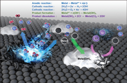
Biodegradable Magnesium Alloys
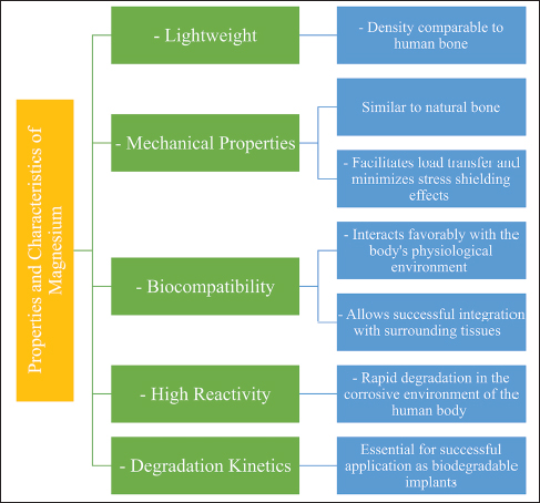
Properties and Characteristics of Pure Magnesium
The key properties and characteristics of pure magnesium as a base material for biodegradable alloys and composites are summarized in Figure 2. Magnesium, with a density of approximately 1.74 g/cm3, closely matches that of human cortical bone (1.8-2.1 g/cm3), enabling effective load distribution and minimising stress shielding effects (Li et al., 2020; Liu et al., 2018). Its elastic modulus (40-45 GPa) is also comparable to natural bone, further reducing the risk of bone resorption (Li et al., 2020). Magnesium exhibits excellent biocompatibility and supports tissue integration without eliciting detrimental responses (Ding et al., 2014; Liu et al., 2018). However, pure magnesium undergoes rapid degradation in chloride-rich physiological fluids (pH 7.4-7.6), releasing hydrogen gas and leading to localised pH changes (Agarwal et al., 2016; Persaud-Sharma & McGoron, 2012; Virtanen, 2011; Witte, 2010).
Benefits and Challenges of Pure Magnesium as a Biodegradable Material
Figure 3 outlines the principal benefits and challenges of pure magnesium as a biodegradable implant material and highlights the need for alloying and reinforcement to overcome its limitations. One notable benefit of pure magnesium lies in its capacity to undergo slow degradation over time, aligning with the healing pace of surrounding tissue (Sezer et al., 2021). This process eliminates the need for secondary surgery to remove the implant, thereby reducing patient discomfort and associated complications (Liu et al., 2018). Moreover, the inherent degradation of magnesium facilitates bone remodelling and tissue regeneration, leading to improved long-term outcomes (Shan et al., 2022).
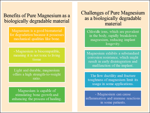
However, the rapid degradation of pure magnesium poses significant challenges for many applications, necessitating alloying and composite reinforcement strategies to control degradation rates and enhance mechanical properties (Amukarimi & Mozafari, 2021; Chandra & Pandey, 2022; Wang et al., 2022). Localised release of magnesium ions may also influence cellular responses and potentially cause cytotoxicity or inflammation if not properly managed (Abdalla et al., 2020; Ben Amara et al., 2023; Venkateswarlu et al., 2024; Zhang et al., 2009).
The Effect of Alloying on Deterioration
The addition of alloying elements such as zinc (Zn), calcium (Ca), strontium (Sr), and manganese (Mn) to magnesium significantly improves both corrosion resistance (stability) and mechanical strength, making magnesium alloys and Mg-MMCs suitable for load-bearing biomedical applications that require structural integrity during the healing period (Aikin et al., 2025; Chen et al., 2019; Kumar & Katyal, 2022; Li et al., 2021; Sharma et al., 2022; Tian & Liu, 2015). For example, Zn and Ca additions refine grain structure, enhance tensile and compressive strength, and slow degradation rates, while Sr at about 3 wt % further refines grains and supports bioactivity, thereby providing adequate mechanical support while minimising premature failure. Careful selection of alloying elements is essential, as some may introduce cytotoxicity concerns or alter degradation behaviour unfavorably. Therefore, a balanced approach is required to optimize these materials for specific implant applications such as bone fixation devices and cardiovascular stents. The major magnesium alloy systems commonly investigated for biomedical applications are illustrated in Figure 4. The influence of common alloying elements on the mechanical properties of biodegradable magnesium alloys and magnesium-based metal matrix composites is summarized in Table 1.
| Sl. No. | Elements | Role in Improving Mechanical Properties |
| 1 | Calcium (Ca) | Enhances grain refinement; increases tensile strength and creep resistance; promotes formation of protective intermetallic phases |
| 2 | Lithium (Li) | Improves ductility while reducing tensile strength; enhances formability in Mg-Li alloys |
| 3 | Zinc (Zn) | Strengthens the composite with optimal enhancement up to 5 wt %; improves tensile and compressive strength |
| 4 | Strontium (Sr) | Improves strength and refines grain size at ~3 wt % addition; enhances bioactivity |
| 5 | Manganese (Mn) | Improves grain fineness and tensile strength |
| 6 | Tin (Sn) | Improves both tensile and compressive strength |
| 7 | Zirconium (Zr) | Provides high damping capacity, tensile strength, and ductility. |
| 8 | Aluminium (Al) | Increases hardness and tensile strength (commonly used in AZ series) |
| 9 | Titanium (Ti) | Improves elongation |
| 10 | Copper (Cu) | Reduces ductility |
| 11 | Silicon (Si) | Enhances fluidity during casting |
| 12 | Iron (Fe) | Not suitable for corrosion resistance; generally avoided |
| 13 | Neodymium (Nd) | Enhances strength (rare earth element) |
| 14 | Molybdenum (Mo) | Increases hardness, Young’s modulus, and elongation |
| 15 | Cerium (Ce) | Reduces yield strength |
Source: Agarwal et al. (2016), Aikin et al. (2025), Chen et al. (2019), Kumar and Katyal (2022), Li et al. (2021), and Sharma et al. (2022).

Magnesium-based Metal Matrix Composite Synthesis and Reinforcement Materials
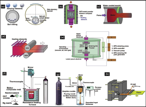
The production of Mg-MMCs can be accomplished through various processing routes. These methods are generally classified into liquid-phase, solid-phase, in situ, deposition, and liquid-solid two-phase techniques (Dash et al., 2018). These metallurgical processing routes are schematically illustrated in Figure 8. Among these, powder metallurgy, friction stir processing, and in situ synthesis are the most commonly used for biomedical Mg-MMCs due to their ability to provide good microstructural control and interfacial bonding.
Powder Metallurgy Techniques
Powder metallurgy is a widely used solid-state method for producing Mg-MMCs. It involves blending magnesium powder with reinforcements (e.g., TiB-, SiC, HA), followed by compaction and sintering. This technique enables uniform distribution of reinforcements and minimizes contamination (Anbuchezhiyan et al., 2015; Dinesh Kumar et al., 2020; Jayasathyakawin et al., 2020; Jiang et al., 2005; Liu et al., 1994; Purohit et al., 2018; Saxena et al., 2023; Wang et al., 2004).
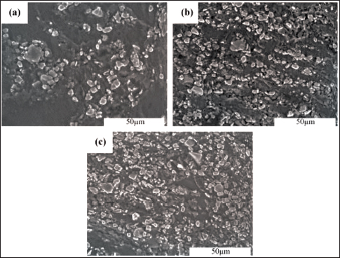
Figure 5 shows scanning electron micrographs of TiB₂/Mg composites. Minor microporosity increases with higher reinforcement volume fractions, with the 20 vol % composite showing the lowest porosity under optimized conditions (Dinesh Kumar et al., 2020).
Friction Stir Processing Techniques
Friction stir processing is a solid-state technique that offers superior microstructural uniformity and strong interfacial bonding without melting the matrix. The friction stir processing technique is cost-effective and environmentally sustainable because it eliminates the need for supplementary binders or consumable materials, which reduces waste generation and energy usage. The lack of molten material throughout the processing phase also mitigates the potential for flaws or inclusions in the ultimate composite. These benefits make friction stir processing an attractive choice for producing magnesium composites with microstructural uniformity for biological and engineering applications. Additional research is still needed to optimize processing settings and to understand how different reinforcements affect friction-stir-processed magnesium composites for biomedical Mg-MMCs (Arokiasamy & Anand Ronald, 2018; Bajakke et al., 2019; Balakrishnan et al., 2015; Dinaharan & Akinlabi, 2018; Kumar et al., 2021; Kundurti et al., 2022; Qiao et al., 2021; Ratna Sunil et al., 2014; Sunil et al., 2016; Vedabouriswaran & Aravindan, 2018).
In Situ Fabrication Methods
In situ synthesis involves direct formation of reinforcements during processing, leading to excellent interfacial bonding and improved corrosion resistance. These methods produce composites with excellent interfacial bonding between the magnesium matrix and the reinforcement phase, including interpenetrating networks formed by reactive infiltration. One common in situ technique is in situ powder metallurgy, where precursor powders of magnesium and reinforcement materials are combined and then processed under controlled conditions to create the composite. Another approach is the in situ solidification method, where the reinforcement materials are introduced to the molten magnesium alloy, and the composite is formed upon cooling. Because in situ fabrication procedures lead to greater bonding between the phases, corrosion resistance and mechanical properties are both improved. However, precise control of the in situ process and potential challenges associated with the inhomogeneous distribution of reinforcement materials require careful consideration during fabrication (Chen et al., 2005; Dong et al., 2004; Meher et al., 2021; Shakil et al., 2020; Shamekh et al., 2012; Wang et al., 2003; Zhou et al., 2022).
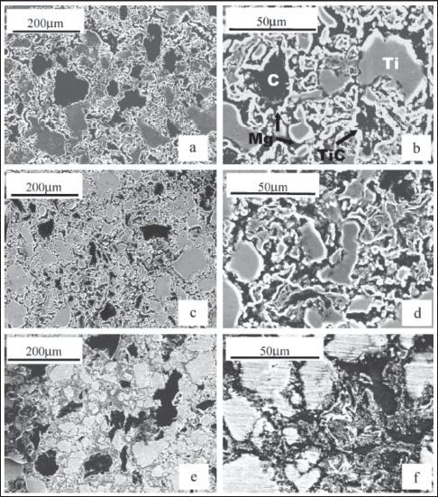
Figure 6 shows the scanning electron micrographs of in situ synthesized Mg matrix composites via reactive infiltration at different temperatures (Dong et al., 2004). The figures demonstrate that reaction completeness and microstructure compactness depend strongly on processing temperature and reactant particle size. Lower porosity and better TiC formation are achieved at optimal temperatures (e.g., 1,023 K), highlighting the importance of process control for biomedical applications.
A comparison of these methods reveals that powder metallurgy offers good scalability, friction stir processing provides excellent grain refinement, and in situ routes yield superior bonding. Selection depends on the target reinforcement type and desired implant performance.
Reinforcement Materials
Ceramics, polymers, and others: Reinforcement materials are critical for improving the mechanical performance and biodegradation behaviour of composites with a magnesium metal matrix (Nie et al., 2021; Su et al., 2020). Commonly used reinforcement materials include ceramics (e.g., calcium phosphate, HA) and polymers (e.g., polycaprolactone, PLGA) (Bommala et al., 2019; Khorashadizade et al., 2021; Kuśnierczyk & Basista, 2017; Mohanasundaram et al., 2023; Ratna Sunil et al., 2014). Ceramics provide excellent biocompatibility and help promote bone regeneration, while polymers offer better toughness and flexibility. In order to ensure that the materials are suitable for the intended implant applications, biocompatibility, rate of degradation, and mechanical qualities are taken into consideration (Cerqueira et al., 2021; Kim et al., 2020). The integration of reinforcing materials leads to enhanced mechanical characteristics, including wear resistance, increased strength, and modulus, thereby rendering the metal matrix composites (MMCs) acceptable for applications involving load-bearing implants. In spite of this, it is essential to optimize the volume fraction and distribution of reinforcement materials in order to prevent agglomeration and keep the composite’s intended attributes (Khorashadizade et al., 2021; Krishnan et al., 2022; Nirala et al., 2021; Ratna Sunil et al., 2014; Su et al., 2020).
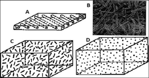
Table 2 summarises commonly used reinforcement materials and their effects on mechanical properties, corrosion resistance, and biocompatibility. Dispersed fibres or particles. Reinforcement materials are typically classified into four main categories: continuous fibres, discontinuous fibers, whiskers, and particulates (Figure 7). The choice of reinforcement materials in MMCs depends on the intended composite properties. The way in which the reinforcement and matrix phases interact is essential for optimizing features such as load transfer, wetting behaviour, reactions, and electrical conductivity. MMCs include reinforcement materials such as ceramics (e.g., SiC and Al2O3), graphite, CNT, graphene, carbon, diamond, cubic boron nitride, and fly ash. Continuous fibres and whiskers are used as uninterrupted reinforcements, whereas discontinuous fibres and particulates act as fragmented reinforcements. In order to obtain the desired outcomes with MMCs, it is necessary to match and blend the reinforcement material with the matrix material in an effective manner (N. Kumar & Soren, 2020).
| Reinforcement Material | Effect on Mechanical Characteristics | Effect on Corrosion Resistance | Biocompatibility |
| SiC | Increases the strength as well as the stiffness and resistance to wear | Improves corrosion resistance | Good biocompatibility |
| Al2O3 | Increases the strength as well as the stiffness and resistance to wear | Improves corrosion resistance | Good biocompatibility |
| TiC | Increases the strength as well as the stiffness and resistance to wear | Improves corrosion resistance | Good biocompatibility |
| BN | Increases the strength as well as the stiffness and resistance to wear | Poor corrosion resistance | Good biocompatibility |
| ZrO2 | Increases the strength as well as the stiffness and resistance to wear | Improves corrosion resistance | Good biocompatibility |
| Hydroxyapatite (HA) | Improves bone-bonding properties | Poor corrosion resistance | Excellent biocompatibility |
| Tricalcium phosphate (TCP) | Improves bone-bonding properties | Poor corrosion resistance | Excellent biocompatibility |
Source: Cerqueira et al. (2021), Khorashadizade et al. (2021), Krishnan et al. (2022), Kuśnierczyk and Basista (2017), Mohanasundaram et al. (2023), Nie et al. (2021), and Su et al. (2020).
Surface Modification Techniques for Enhanced Biocompatibility
Increasing the biocompatibility and bioactivity of Mg-MMCs can be accomplished through the application of surface modification techniques (Delavar et al., 2023). In order to promote cell adhesion and aid osseointegration, the surface of the composite may be modified or coated with bioactive compounds such as HA or biodegradable polymers (Buck et al., 2020). Surface modification has a dual role in improving the initial cellular response and mitigating the release of corrosion by-products, hence minimising potential harmful effects on the surrounding tissues. In addition, the application of surface modification techniques can accomplish effective control of the rate of degradation of composites and make it possible for the regulated release of magnesium ions. This approach serves to mitigate the potential cytotoxic effects associated with the material. Surface modification is frequently performed using techniques such as sol-gel coating, plasma electrolytic oxidation (PEO), plasma spraying, and electrospinning to support controlled ion release and to delay excessive hydrogen gas evolution. Optimizing surface characteristics in magnesium composites is crucial to enhance biocompatibility and ensure the long-term success of implants for biomedical applications (da Conceicao et al., 2010; Daroonparvar et al., 2022; Yang et al., 2011).
Mechanical Performance of Magnesium Composites
Strength in Tension, Strength in Yield, and Ductility
Parameters such as tensile strength, ductility, and yield strength are vital when determining whether magnesium composites are suitable for use as biomedical implants. The mechanical performance of magnesium composites is characterised by parameters such as these. Tensile strength denotes the highest stress that a material can withstand without breaking under tension. The yield strength indicates the stress value at which plastic deformation commences, providing insight into the material’s capacity to resist permanent deformation. Ductility describes the degree to which a substance can deform plastically before it fractures. The mechanical properties of magnesium composites are susceptible to being significantly influenced by the reinforcement materials used. It is crucial to keep in mind that the addition of ceramics or polymers, for example, has the potential to raise both the tensile strength and the yield strength of the material; nevertheless, it is important to keep in mind that this may come at the expense of decreased ductility in the final product. The attainment of a harmonious equilibrium among these characteristics is of utmost importance in order to guarantee the composite’s ability to endure mechanical stresses without untimely breakdown, while simultaneously permitting sufficient flexibility to accommodate physiological motions (Haghshenas, 2017; Khorashadizade et al., 2021; Krishnan et al., 2022; Li et al., 2014; Venugopal et al., 2023; Zhang et al., 2009).
Figure 9 illustrates the tensile strength and elongation of Mg-Zn-Mn alloys in their respective as-cast and extruded forms. When looking at the tensile strengths of the material, such as the yield strength and ultimate tensile strength (UTS), Figure 9(a) demonstrates that the tensile strength (both yield strength and ultimate tensile strength) of Mg-Zn-Mn alloys increases with increasing Zn content. The extrusion process results in a substantial enhancement in tensile properties compared to the as-cast condition. For instance, the yield strength of the Mg-1Zn-1Mn alloy increased from 44 MPa (as-cast) to 246 MPa after extrusion (a 459% improvement), while the ultimate tensile strength rose from 174 MPa to 280 MPa (a 60% improvement) (Zhang et al., 2009).
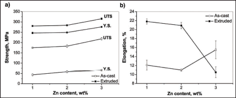
Figure 9(b) shows the elongation behaviour of Mg-Zn-Mn alloys. Although increasing Zn content slightly improves elongation in the as-cast alloys, the elongation of extruded alloys decreases significantly at higher Zn levels. For the Mg-Zn-Mn alloy, the elongation drops to approximately 10% after extrusion, which is lower than that of the corresponding as-cast alloy (Zhang et al., 2009). This trade-off between strength and ductility must be carefully considered when designing load-bearing biomedical implants.
Hardness and Wear Resistance
Magnesium composites’ mechanical properties, particularly their hardness and wear resistance, are critical in determining the longevity and effectiveness of these materials when applied as biomedical implants. The term “hardness” pertains to the capacity of a material to resist deformation and penetration, whereas “wear resistance” denotes its ability to endure abrasion and surface degradation caused by frictional forces. The inclusion of reinforcement materials, such as ceramics, has the prospective to greatly improve hardness and wear resistance, hence rendering the composite more suited for applications that bear loads. Nevertheless, the utilization of an implant that is too rigid might result in heightened abrasion on surfaces that come into contact with it. This necessitates the need for a meticulous equilibrium to be established, in order to attain ideal performance in terms of wear, while simultaneously avoiding excessive wear on natural tissues. To establish which applications are best suited for composites and to assure the continuous success of implants, it is critically necessary to have a full understanding of the hardness and wear parameters of composites (Alahelisten et al., 1993; Madhusudhan Reddy et al., 2013; Soorya Prakash et al., 2016; Wang et al., 2004, 2006).
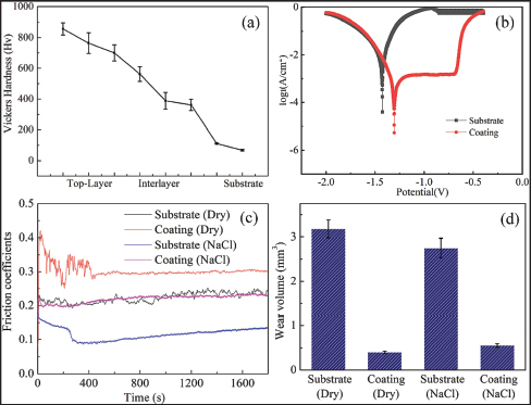
Figure 10a illustrates how the Vickers hardness varies with depth in the graded coatings. It demonstrates a steady drop in average hardness from the top layer (856.5 HV) to the substrate (67.8 HV) over the course of the coating. Because WC particle doping had good effects on the Ni-WC composite coatings, the top layer’s hardness was noticeably approximately 12.6 times larger than that of the substrate. This doping increases hardness by dispersing and strengthening the fine grain. Figure 10b illustrates the potentiodynamic polarisation curves for both the coating and the substrate, respectively. The results show that the coating has a higher corrosion potential and lower corrosion current density than the substrate. This indicates improved corrosion resistance. Figure 10c and 10d illustrates the coefficient of friction (COF) and wear behaviour, respectively. The coatings had a higher COF than the Mg alloy because of the rough surface of the coating. The top layer’s matrix was worn away first, leaving robust WC particles on the surface during wear testing. This resulted in a rough coating surface and an elevated COF. Despite that, the coating showed a substantial decrease of 87.4% and 80.0% in wear volumes under dry and NaCl circumstances, showing about five to eight times higher wear resistance compared to the Mg alloy (Wang et al., 2023).
Behaviour Caused by Fatigue and Breaking Toughness
The investigation of fatigue characteristics and fracture toughness is of utmost importance when considering the mechanical dependability and safety of magnesium composites in dynamic load-bearing circumstances. The term “fatigue behaviour” refers to the way a composite material behaves when subjected to the influence of cyclic loading and unloading, which has the potential to cause the material to fail due to fatigue over the course of some amount of time. The inclusion of reinforcement materials has the potential to improve the fatigue resistance of a material by increasing the material’s resistance to cracking and reducing the amount of stress concentrated in certain areas of the material. This enhancement is possible as a result of the addition of materials that provide reinforcement to the structure. The fracture toughness parameter quantifies the material’s capacity to resist the propagation of cracks, hence reflecting its capability to endure abrupt impacts or stress concentrations without experiencing catastrophic collapse. A composite material possessing a high fracture toughness exhibits enhanced resistance to unanticipated mechanical stresses encountered during the utilization of implants. The comprehension of fatigue characteristics and fracture toughness of magnesium composites enables researchers and engineers to develop implants capable of withstanding physiological loads for prolonged durations while simultaneously reducing the likelihood of abrupt failure and related consequences. The accurate evaluation and optimisation of the mechanical characteristics of magnesium composites are crucial in guaranteeing the sustained performance and dependability of these materials in biomedical implant applications (García-Romero et al., 2010; Hack et al., 1984; Qi et al., 2017; Shin & Bae, 2018; Vaidya & Lewandowski, 1996; Xia et al., 2020; Zhu & Iizuka, 2003).
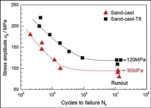
The S-N curves for sand-cast and sand-cast-T6 Mg-10Gd-3Y-0.5Zr alloys that were subjected to rotating beam loading (with a stress ratio of -1) at about 100 Hz in an air environment are displayed in Figure 11. These alloys were loaded with a stress ratio of -1, and the frequency of the rotating beam loading was approximately 100 Hz. The resistance to fatigue of these alloys is under examination. The fatigue strength of the sand-cast Mg-10Gd-3Y-0.5Zr alloy was found to be 95 MPa after 107 cycles, but the sand-cast-T6 Mg-10Gd-3Y-0.5Zr alloy demonstrated a fatigue strength of 120 MPa for the same cycle count. This was established after comparing the two alloys’ fatigue strengths. The application of the T6 thermal treatment process resulted in a 26% increase in the end product’s fatigue strength (Liu et al., 2014).
Corrosion Behaviour of Magnesium-based Composites
Magnesium Degradation Mechanisms in Physiological Environments
The comprehension of the corrosion characteristics exhibited by magnesium-based composites is of utmost importance in order to effectively utilize them as biodegradable materials for biomedical implants. Magnesium degrades through electrochemical mechanisms upon exposure to the physiological environment. This degradation is mostly attributed to the relationship between magnesium and water, as well as the presence of ions in bodily fluids. One of the stages of deterioration is the dissolution of magnesium, which leads to the creation of magnesium ions and ultimately ends in the emission of hydrogen gas. The aforementioned procedure has the capability to induce localised alterations in pH levels, which may result in either alkalisation or acidification in the immediate area of the implant. These changes have the capacity to impact the surrounding tissues and cells. In addition, the liberation of magnesium ions could potentially exert physiological influences on cellular mechanisms and tissue reactions. Several factors, including alloy composition, microstructure, surface area, and exposure circumstances, contribute to the deterioration rate. Understanding these degradation mechanisms comprehensively is extremely important to accurately forecast the performance of the material and customise its degradation rate in line with the healing process for specific biomedical implant applications (Bakhsheshi-Rad et al., 2014, 2016; Eliezer & Witte, 2010; Neves et al., 2021; Pulido-González et al., 2020; Razavi et al., 2010; Törne et al., 2017; Xu et al., 2022; Zhang et al., 2013).
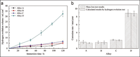
The results of studies on hydrogen evolution and mass loss are depicted in Figure 12, which includes hydrogen evolution curves (Figure 12a) and corrosion rates of alloys (Figure 12b). These results reveal distinct corrosion behaviours. After 120 h of immersion, Alloy A’s hydrogen evolution volume aligns with Alloy B’s, while Alloy C displays elevated hydrogen evolution, and Alloy D’s mean volume surpasses those of Alloys A and B by sixfold. The results of mass loss testing indicate that Alloy B has a slightly lower corrosion rate than Alloy A. However, Alloy C and Alloy D have corrosion rates that are 32% and 437% higher, respectively, than Alloy A. Despite the fact that the rates acquired using this approach are slightly lower than those obtained using the mass loss tests, the findings of the hydrogen evolution tests support these findings and reveal a trend that is comparable. This is the case despite the fact that the rates obtained using this method are lower than those obtained using the mass loss tests. This is due to the fact that during the process of simulated body fluids (SBF) renewal, there is a chance that hydrogen may be released into the air and then dissolved into SBF. Remarkably, Figure 12 showcases a direct relationship between Ag content and corrosion rate increase, notably with Alloy B (0.2 wt% Ag) mirroring the rate of Ag-free alloy. These corrosion behaviours are influenced by grain size, second-phase characteristics, distribution, and the presence of the MgZr compound, with finer grains generally improving corrosion resistance while discontinuous second phases enhance microgalvanic-driven corrosion effects (Zhang et al., 2013).
Corrosion Protection Strategies
The topics of interest include alloying, coatings, and surface treatments. In order to mitigate the issues arising from the quick deterioration of pure magnesium, many solutions for corrosion protection are implemented in magnesium-based composites. Alloying is a frequently used technique that involves introducing specific elements into the magnesium matrix to modify its corrosion characteristics and regulate the rate of degradation (Abdalla et al., 2020; Heimann, 2021). The addition of alloying metals such as aluminium, calcium, or zinc has the potential to generate protective corrosion layers or compounds. This, in turn, can effectively decrease the rate at which magnesium ions are released and enhance the overall resistance to corrosion (Agarwal et al., 2016; Aikin et al., 2025; Delavar et al., 2023; Virtanen, 2011). A further approach entails the application of protective coatings or surface treatments to the composite material. Coatings composed of bioceramic materials, such as HA, or biodegradable polymers have the ability to establish a protective barrier between the composite material and the surrounding physiological environment. This barrier serves to decelerate the pace of degradation and improve the biocompatibility of the composite (Ratna Sunil et al., 2014). In addition, the application of surface treatments such as anodization or PEO can result in the manufacture of coatings on the surface of the composite material that are long-lasting and resistant to corrosion. These coatings can be advantageous in a variety of applications. The comprehension and enhancement of these corrosion mitigation approaches are of utmost importance in guaranteeing the enduring stability and efficacy of magnesium-based composites employed in biomedical implantation contexts (Allavikutty et al., 2021; Heimann, 2021; Li & Zheng, 2013; Narayanan et al., 2015; Yang et al., 2010).
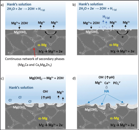
Figure 13 depicts the mechanism of corrosion that takes place in the Mg-Zn-Ca alloy after it has been cast. This mechanism of corrosion takes place when the resulting alloy is immersed in Hank’s solution. The first steps in the process involve the creation of a protective layer and the breakdown of the alloy in a manner that is consistent throughout (Figure 13a), but local disruption of the layer by chlorine ions and hydrogen bubbles leads to the formation of small pits that expose alloy portions to the electrolyte (Figure 13b). The galvanic interaction between the α-Mg matrix and the intermetallic secondary phases also contributes to the acceleration of this process. The presence of the continuous phase at the grain boundary slows corrosion advancement (Figure 13c), even though the intermetallic phase itself does not disintegrate. As dissolution continues, corrosion progresses through the network of secondary phases (Figure 13d) and finally through the α-Mg matrix via localised corrosion (Figure 13c), exacerbating the overall corrosion. The production of HA is triggered on the residual protective Mg(OH)2 layer as a result of the interaction between free Mg2+ and Ca2+ cations and solution phosphates (Figure 13d). This formation is made easier by the rise in pH that results from the presence of OH- in the solution (Pulido-González et al., 2020).
In Vitro and In Vivo Corrosion Studies
Comprehensive corrosion investigations are carried out both in the laboratory (in the form of in vitro testing) and inside living creatures (in the form of in vivo testing) in order to evaluate the corrosion properties and biocompatibility of magnesium-based composites. In vitro investigations entail submerging the composite specimens in simulated bodily fluid or cell culture conditions to replicate the physiological milieu and evaluate the rate of degradation and kinetics of ion release (Hussein et al., 2022). These findings offer significant insights into the first corrosion behaviour of the material and its possible implications for adjacent cells and tissues (Sommer et al., 2022; Tahriri et al., 2018). The process of in vivo corrosion investigations entails the insertion of composites into animal models for the purpose of assessing their degradation characteristics, tissue reaction, and suitability for biological applications in a setting that closely mimics real-life biological conditions (Sommer et al., 2022). These studies make it possible to monitor the continuously degrading performance of the implanted composite as well as the host’s reaction to the composite. Understanding the biodegradation kinetics, biocompatibility, and safety features of magnesium-based composites requires the combined use of in vitro and in vivo corrosion research. This is of the utmost importance. These researches provide valuable insights that aid in the refinement of these composites, ensuring their secure and efficient utilization across diverse biomedical implant applications (Cho et al., 2022; Chou et al., 2013; Sanchez et al., 2015; Wen et al., 2021).
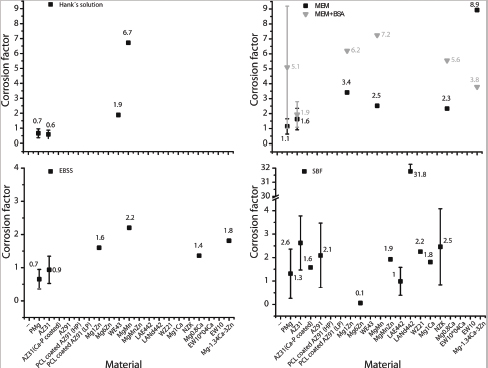
Corrosion media used in in vitro testing, such as Earle’s balanced salt solution (EBSS), SBF, Hanks’ balanced salt solution (HBSS), minimum essential media (MEM), and MEM supplemented with bovine serum albumin (BSA), all have distinct effects on the test specimens, as shown in Figure 14. Importantly, the corrosion factors determined for a given corrosion solution show considerable similarity, with only a few exceptions. In general, the corrosion factors for EBSS and SBF are below 3, while MEM exhibits higher values, which range from 1.5 to 3.5. Upon introduction of proteins, the values increase to 4-7. In contrast, the corrosion factors yielded by Hanks’ solution differ significantly; yet definitive correlation assessment is hindered due to limited statistical data (Sanchez et al., 2015).
Biocompatibility and Biological Response
Interaction of Magnesium Composites with Cells and Tissues
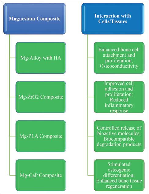
The assessment of biocompatibility in magnesium composites plays a pivotal role in determining their appropriateness for utilization in biomedical implant applications (Huan et al., 2010; Zhang et al., 2009). Upon being inserted into the human body, these composite materials engage in interactions with cells and tissues, thereby beginning biological reactions that have an impact on their integration and overall functionality. The initial stage of the interaction involves the surface of the composite material, where proteins from the surrounding environment adhere and promote the attachment of cells. Various types of cells, including osteoblasts and fibroblasts, engage in interactions with the composite surface, thereby facilitating the processes of tissue growth and remodelling. The degradation products of the composite material, which encompass magnesium ions, are additionally implicated in biological responses. The concentration of magnesium ions can elicit either stimulatory or inhibitory effects on several cellular processes, including proliferation, differentiation, and apoptosis. Gaining a comprehensive understanding of the interplay between magnesium composites and cellular and tissue components is of paramount importance in facilitating desirable tissue reactions and guaranteeing successful incorporation of the implant within the host milieu (Al-Shalawi, Azmah Hanim, et al., 2023; Castro et al., 2019; Huan et al., 2010; Shunmugasamy et al., 2023). Figure 15 summarises the effects of representative magnesium composites on cells and tissues.
Inflammatory Response and Host Reactions
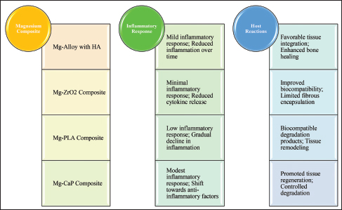
When introduced into the human body, magnesium composites have the potential to elicit an inflammatory response, which is an inherent component of the physiological wound healing process (Al-Shalawi, Azmah Hanim, et al., 2023; Castro et al., 2019; Huan et al., 2010; Li et al., 2022; Shunmugasamy et al., 2023). It is possible to identify the beginning stages of inflammation by the movement of immune cells to the site of implantation. These immune cells may include neutrophils and macrophages. The immune system’s cells perform an essential part in the removal of waste from cells and the regulation of the local microenvironment in the tissue that surrounds them. On the other hand, inflammation that is excessive or persists for an extended period of time can have negative effects on the host, such as the development of chronic inflammation or the production of fibrous encapsulation surrounding the implant. The composite’s rate of degradation can be affected by the inflammatory response, which impacts the local electrolyte balance and pH. The management of degradation rate and the manipulation of surface qualities of composites are crucial factors in mitigating the inflammatory response and facilitating a controlled and positive host reaction towards the implant (Avery et al., 2023; Bakhsheshi-Rad et al., 2017; Hotchkiss et al., 2018; Hua et al., 2023; Huan et al., 2010; Jurczyk et al., 2020; Martinez et al., 2023; Prestat & Thierry, 2021; Saha et al., 2022; Selders et al., 2017; Velnar et al., 2016). Figure 16 summarises magnesium-composite-induced inflammation and the associated host reactions.
Immunomodulatory Effects and Osteogenesis
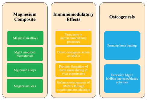
Magnesium composites possess immunomodulatory properties that can exert an influence on the immune response and subsequent tissue healing processes (He et al., 2023). The composite material has the ability to regulate immune cell activity and cytokine production through the release of magnesium ions. This has an impact on the equilibrium between anti-inflammatory and pro-inflammatory responses (Zhou et al., 2021). The appropriate modulation of the immune system can effectively facilitate the process of tissue repair and regeneration, while concurrently mitigating the occurrence of unfavourable reactions. Moreover, magnesium composites have demonstrated potential in facilitating osteogenesis, which is the biological process responsible for bone formation (He et al., 2023). Magnesium ions released during degradation can promote osteogenic differentiation of mesenchymal stem cells, which supports bone regeneration and stronger bonding between the implant and adjacent bone tissue (He et al., 2023; Zhou et al., 2021). Additionally, the similarity between magnesium and natural bone minerals can create a conducive microenvironment for the functioning of osteoblasts and the creation of the extracellular matrix (He et al., 2023). Figure 17 schematically presents the immunomodulatory effects and osteogenic potential of magnesium composites. It is of utmost importance to comprehend the immunomodulatory effects and osteogenic capacity of magnesium composites in order to enhance their biocompatibility and guarantee the successful integration and usefulness of these materials in biomedical implant applications connected to bone (Loi et al., 2016; Medhat et al., 2019; Xue et al., 2022; Zhao et al., 2022).
Preclinical and Clinical Studies
Preclinical Evaluation of Magnesium-based Implants
Prior to conducting human trials, magnesium-based implants undergo thorough preclinical examination in laboratory settings. This stage evaluates implant biocompatibility, mechanical characteristics, and degradation patterns in vitro and ex vivo (Chagnon et al., 2019). In vitro investigations encompass the exposure of implants to simulated bodily fluids or cell cultures, enabling the examination of their interactions with biological constituents (Hua et al., 2023; Huan et al., 2010; Loi et al., 2016). The degradation rate, release of magnesium ions, and cellular reactions of the implants are meticulously examined in order to comprehend their behaviour within a controlled setting (Chen et al., 2019; Li et al., 2014; Nene et al., 2016; Shan et al., 2022). Furthermore, ex vivo tests may encompass the transplantation of the composites into animal models’ tissues or organs that exhibit a high degree of similarity to human physiology (Bakhsheshi-Rad et al., 2017; Li et al., 2022; Martinez et al., 2023; Sanchez et al., 2015; Sommer et al., 2022). These experiments offer significant insights into the early biological reaction and degrading behaviour of the implants within a scenario that closely resembles real-world conditions. The preclinical assessment is a critical step in identifying potential problems, improving implant designs, and predicting their performance before proceeding to animal testing and clinical trials (Chagnon et al., 2019; Mathew et al., 2023; Sekar et al., 2021).
Animal Studies: Efficacy and Safety Assessment
The examination of animals is an essential component in the assessment of the effectiveness and safety of magnesium-based implants within living beings. These investigations entail the insertion of the composites into animal models, commonly including rabbits or pigs, in order to evaluate their efficacy inside a functioning biological system (Sun et al., 2021). Table 3 summarises the outcomes of an initial minipig study of magnesium implants, organised by implantation site, together with the associated safety evaluations (Imwinkelried et al., 2020). The present investigation examines implant biocompatibility, deterioration, tissue integration, and host reactions (Rahim et al., 2020; Sun et al., 2021). The implants were monitored over a prolonged duration in order to evaluate the enduring consequences and possible problems. Imaging modalities, histopathological examination, and biomechanical assessment are often employed methodologies for the evaluation of implant efficacy and tissue reactions (Grün et al., 2018). Animal research plays a crucial role in gathering essential information on the biological processes, tissue regeneration, and possible negative effects of implants. The aforementioned findings provide valuable insights for enhancing the design and safety of implants prior to embarking on human clinical trials (Grün et al., 2018; Rahim et al., 2020; Sekar et al., 2021; Sun et al., 2021).
| Conditions Required for Implantation | Situation of Degradation/Loading | Safety Considerations | Results | References |
| Nasal bone: Tissue pocket with rectangular plate | Degradation without load | Consistency of degradation | Limited localised corrosion | |
| Maintenance of strength | The initial bending strength is at 80% after 12 weeks | |||
| Gas release | Coating prevents initial peak release | |||
| Frontal bone: Contoured plates using non-locking screws | The plates are against the bone and the screws tightened for tension | Screw head rupture during implantation | Replaceable over drilled Screws | |
| Gas release | Dependent on screw location | |||
| Plate–screw connection | No preferential degradation observed | |||
| Mandible: Thin-walled rivet-screws for cortical bone | Implant deformed plastically while undergoing load | Effects of plastic deformation and loading | Coating delays gas release despite deformation | |
| Rib arches: Contoured plates using non-locking screws | Bone plates are fitted and screws are tightened to move the ribs in a partial osteotomy | Effect of respiration and animal body weight | One pilot animal experienced a broken Mg mandible plate | |
| The rib-specific plates are intact, but some screws are broken | ||||
| Systemic effects: Combined Implants | Total degradation products | Buildup of metals in body organs due to alloying | No accumulation except for Zr | |
| Release of gas | No systemic effects observed |
Clinical Trials and Case Studies of Magnesium-based Implants
Clinical studies provide the final assessment of magnesium-based implants’ safety and efficacy in humans (Zhao et al., 2017). The trials encompass meticulously planned investigations wherein human subjects are administered implants within controlled environments (Sekar et al., 2021). The primary objectives of clinical trials include assessing the implants’ biocompatibility, degradation kinetics, mechanical performance, and long-term outcomes in real-world scenarios (Prestat & Thierry, 2021). The trials are typically conducted in phases, starting with small groups to evaluate safety, followed by larger groups to assess efficacy and gather statistical data. Case studies may also be conducted to report individual patient experiences and outcomes with magnesium-based implants. The data from clinical trials and case studies help establish the safety profile and effectiveness of the implants in human patients. Positive results from clinical trials pave the way for regulatory approval and widespread clinical use of magnesium-based implants in specific medical applications (Albaraghtheh et al., 2022; Espiritu et al., 2021, 2022; Garimella et al., 2026; Jurczyk et al., 2020; Prestat & Thierry, 2021; Sekar et al., 2021; Stürznickel et al., 2021; Suljevic et al., 2022; Venkateswarlu et al., 2023; Zhao et al., 2017).
Applications of Magnesium Metal Matrix Composites in Biomedical Implants
Bone Fixation Devices and Osteosynthesis
Mg-MMCs find application in bone fixation devices and osteosynthesis, where they play a crucial role in stabilizing fractured bones and supporting the healing process (Xue et al., 2022). These implants are used in orthopaedic surgeries to immobilise fractured bones and facilitate bone fusion. The biodegradability of magnesium composites is particularly helpful in this context because, as they gradually disintegrate over time, they lower the risk of implant-associated problems and eliminate the requirement for a further operation for removal (Medhat et al., 2019; Xue et al., 2022; Zhao et al., 2022). Furthermore, the mechanical properties of magnesium composites are remarkably close to those of genuine bone. This promotes load transfer throughout the healing process and lessens the effects of stress shielding. The controlled degradation of magnesium composites in bone fixation devices can stimulate bone formation and remodelling, leading to improved long-term outcomes. In order to ensure an adequate level of implant integrity during the entire healing period, the rate of deterioration and the mechanical properties of the composite are carefully examined. Bone fixation devices and osteosynthesis with magnesium composites represent a promising area for enhancing patient outcomes and reducing the burden of implant-related surgeries (Cerqueira et al., 2021; Espiritu et al., 2022; Fu et al., 2011; Garimella et al., 2026; Kim et al., 2020; Munir et al., 2023; Putra et al., 2020; Tahriri et al., 2018; Zhang et al., 2021; Zhou et al., 2021).
Cardiovascular Stents and Vascular Applications
The investigation of magnesium-based composites for cardiovascular stents and vascular applications is now underway (Walker et al., 2014). Stents are cylindrical instruments employed for the management of coronary artery disease and various vascular blockages (Ahadi et al., 2023). The utilization of magnesium composites in stents presents numerous benefits, including the provision of temporary scaffolding support throughout the healing process and slow degradation, thereby obviating the necessity for permanent implants (Erbel et al., 2007). Ensuring the biocompatibility of magnesium composites is of utmost importance in mitigating the potential for inflammation and thrombosis commonly associated with conventional stents (Ahadi et al., 2023). Magnesium composites, when intentionally degraded for use in vascular applications, may be able to solve the problem of in-stent restenosis that plagues conventional metal stents (Ma et al., 2016). Nevertheless, it is crucial to optimize the degradation kinetics, mechanical strength, and surface qualities in order to guarantee the stent’s mechanical support throughout the healing process and mitigate the risk of premature stent failure. Ongoing research in this domain demonstrates the potential of magnesium composites to significantly transform the field of cardiovascular treatments by providing enhanced biocompatibility and patient-centric solutions (Erbel et al., 2007; Ma et al., 2016; Walker et al., 2014).
Orthopaedic Implants and Soft Tissue Scaffolds
Mg-MMCs exhibit considerable potential for application in diverse orthopaedic implant and soft tissue scaffold designs (He et al., 2023). Within the field of orthopaedics, these composite materials find application in several types of implants, including spinal cages, interbody fusion devices, and craniomaxillofacial plates (Kim et al., 2018; Schaller, Saulacic, Imwinkelried, et al., 2016; Shen et al., 2019). The favourable nature of magnesium composites lies in their biodegradability, which proves beneficial in various applications. Specifically, these implants offer support during the healing process while concurrently undergoing slow degradation as bone tissue undergoes regeneration (Medhat et al., 2019). It is possible to tailor the composite material’s mechanical properties to meet the specific requirements of orthopaedic applications; this results in a solution that is both adaptable and individualized for patients (Chandra & Pandey, 2020; Tsakiris et al., 2021; Walker et al., 2014). In addition to this, magnesium composites have the potential to function as scaffolds in the field of tissue engineering and regenerative medicine (Sheikh et al., 2015; Xue et al., 2022). Temporary frameworks are utilized to facilitate tissue growth and remodelling processes by stimulating the development of new tissue (Adetunla et al., 2022; Cerqueira et al., 2021; Huang et al., 2014; Persaud-Sharma & McGoron, 2012). The progressive deterioration of the composite material facilitates the development of a functional tissue architecture, leading to the eventual replacement of the implant by native tissue. However, in order to guarantee the efficiency and safety of these implants and scaffolds in the context of orthopaedic and tissue engineering activities, it is absolutely necessary to conduct a comprehensive analysis of the rate of degradation, the mechanical features, and the biocompatibility of the material (Adetunla et al., 2022; Huang et al., 2014; Radha & Sreekanth, 2017; Rahman et al., 2020).
Challenges and Future Perspectives
Biodegradation Control and Implant Longevity
The management of the biodegradation rate of magnesium-based composites is one of the key challenges that arises in the process of utilizing these materials for the manufacture of biomedical implants (Rahman et al., 2020). The progressive deterioration of implants is advantageous in reducing the necessity for implant extraction procedures. However, it is crucial to strike a balance that guarantees adequate mechanical reinforcement during the entirety of the healing phase (Adetunla et al., 2022; Zhao et al., 2017). Rapid deterioration can cause premature implant failure and excessive hydrogen evolution, while second-phase particles and reinforcements may accelerate attack through microgalvanic coupling; delayed degradation may impede tissue repair (Zhao et al., 2017). The attainment of an optimal degradation rate is contingent upon the meticulous choice of alloying components, composite composition, and surface changes (Aikin et al., 2025). Moreover, the degrading behaviour of implants might be influenced by changes in physiological settings among individuals, necessitating the implementation of individualized approaches in implant design. The maintenance of suitable biodegradation management is of utmost importance in order to attain optimal longevity of implants and favourable outcomes for patients (Adetunla et al., 2022; Zhao et al., 2017).
Cytotoxicity and Biocompatibility Concerns
The release of magnesium ions during the degradation process of magnesium composites has the potential to have an effect on both cytotoxicity and biological responses (Xin et al., 2011). Magnesium ions, when present in low concentrations, have been shown to encourage the growth of new cells and the repair of damaged tissue; however, magnesium ions present in high concentrations have been shown to have the opposite effect, leading to cell death and inflammation (Zhao et al., 2017). Properly tuning the degradation rate and ion release kinetics is essential to minimize cytotoxicity and ensure biocompatibility. Additionally, variations in individual responses to magnesium ions must be considered, as some patients may be more sensitive to their presence. In order to address cytotoxicity and biocompatibility concerns before moving on to clinical applications, it is absolutely necessary to carry out complete biocompatibility assessments. These assessments should include both in vitro and in vivo research (Chagnon et al., 2019; Sezer et al., 2018; Xin et al., 2011).
Translation to Commercial Products and Regulatory Aspects
Despite significant research advancements, the translation of magnesium-based composites into commercial products for widespread clinical use involves various challenges (Walker et al., 2014). Manufacturing scalability, consistent quality control, and process reproducibility remain significant concerns (Wang et al., 2022). Additionally, regulatory aspects related to biodegradable materials and medical devices must be thoroughly addressed to meet safety and efficacy standards for clinical applications. Obtaining regulatory approvals can be a lengthy and resource-intensive process. Collaborations between academic researchers, medical device companies, and regulatory bodies are crucial for navigating these challenges and bringing magnesium-based composites to market. Moreover, long-term post-market surveillance is essential to monitor the performance and safety of these implants in real-world scenarios. In order to realize the full potential of magnesium-based composites as novel and patient-friendly solutions in the field of biomedical implants, it is essential to overcome these hurdles and handle regulatory considerations (Al-Shalawi, Azmah Hanim, et al., 2023; Al-Shalawi, Mohamed Ariff, et al., 2023; Chagnon et al., 2019; Chandra & Pandey, 2020; Drelich et al., 2020; Hung et al., 2019; Prakasam et al., 2017; Sezer et al., 2018; Wang et al., 2022; Wu et al., 2015; Zhao et al., 2017; Zhu et al., 2022).
Conclusion
Because of properties such as biocompatibility, mechanical qualities that are similar to natural bone, and controlled biodegradation, magnesium-based composites provide a compelling alternative to standard nondegradable implants. The properties and characteristics of magnesium, including its lightweight nature and biocompatibility, make it an attractive material for medical applications. Alloying with specific elements allows for the optimisation of degradation kinetics and mechanical performance, tailoring the composites to specific implant requirements. The corrosion behaviour of magnesium composites was explored in this article. The article also emphasized the need for biodegradation control to ensure optimal implant longevity and prevent premature failure. Biocompatibility work continues to flag risks linked to magnesium ion release, so careful material design and thorough biocompatibility evaluation remain necessary. The review article contained a complete analysis of the many applications of magnesium composites in the realm of biomedical implants as well as other applications. These applications cover a wide variety of medical devices, including stents for the circulatory system, orthopaedic implants, bone fixation devices, and scaffolds for soft tissue. The exhibited applications have showcased the potential of magnesium composites in improving patient outcomes and minimising the necessity for subsequent surgeries through the facilitation of progressive degradation and tissue regeneration. When considering the future, the outlook for the utilization of biodegradable magnesium-based composites in biomedical implants is highly favourable. Current research endeavors are focused on tackling the obstacles associated with degradation control, biocompatibility, and regulatory considerations in order to facilitate the successful transition of these innovations into commercially viable products. The establishment of collaborations among academia, industry, and regulatory agencies is of utmost importance in effectively navigating the intricate journey from preclinical assessment to clinical trials and eventual market acceptance. In short, biodegradable magnesium composites remain promising temporary implant materials when mechanical support, biocompatibility, and degradation rate are jointly managed. Continued progress in processing, surface engineering, and clinical evaluation will determine how widely they enter routine medical device practice.
Acknowledgement
The authors also acknowledge the valuable suggestions of Professor (Dr.) Goutam Sutradhar in submitting this manuscript.
Data Availability
No data set is available with the article because no new data were created/analysed in this study.
The authors declared no potential conflicts of interest with respect to the research, authorship, and/or publication of this article.
The authors disclosed receipt of the following financial support for the research, authorship, and/or publication of this article: Anusandhan National Research Foundation (ANRF) under Partnerships for Accelerated Innovation and Research (PAIR) with IIT(ISM) Dhanbad for the project “Development of Innovative and Cutting-Edge Indigenous Technologies for Critical Minerals Exploration and Smart/Sustainable Mining,” Sanction order no. ANRF/PAIR/2025/000027/PAIR-B.
Abdalla M., Joplin A., Elahinia M., & Ibrahim H. (2020). Corrosion modeling of magnesium and its alloys for biomedical applications: Review. Corrosion and Materials Degradation, 1(2), 219–248. https://doi.org/10.3390/cmd1020011
Adetunla A., Fide–Akwuobi A., Benjamin H., Adeyinka A., & Kolawole A. (2022). A study of degradable orthopedic implant: An insight in magnesium metal matrix composites. Heliyon, 8(9), e10503. https://doi.org/10.1016/j.heliyon.2022.e10503
Agarwal S., Curtin J., Duffy B., & Jaiswal S. (2016). Biodegradable magnesium alloys for orthopaedic applications: A review on corrosion, biocompatibility and surface modifications. Materials Science and Engineering: C, 68, 948–963. https://doi.org/10.1016/j.msec.2016.06.020
Ahadi F., Azadi M., Biglari M., Bodaghi M., & Khaleghian A. (2023). Evaluation of coronary stents: A review of types, materials, processing techniques, design, and problems. Heliyon, 9(2), e13575. https://doi.org/10.1016/j.heliyon.2023.e13575
Aikin M., Shalomeev V., Kukhar V., Kostryzhev A., Kuziev I., Kulynych V., Dykha O., Dytyniuk V., Shapoval O., Zagorskis A., Burko V., Khliestova O., Titov V., & Hrushko O. (2025). Recent advances in biodegradable magnesium alloys for medical implants: Evolution, innovations, and clinical translation. Crystals, 15(8), 671. https://doi.org/10.3390/cryst15080671
Alahelisten A., Bergman F., Olsson M., & Hogmark S. (1993). On the wear of aluminium and magnesium metal matrix composites. Wear, 165(2), 221–226. https://doi.org/10.1016/0043–1648(93)90338–M
Albaraghtheh T., Willumeit–Römer R., & Zeller–Plumhoff B. (2022). In silico studies of magnesium–based implants: A review of the current stage and challenges. Journal of Magnesium and Alloys, 10(11), 2968–2996. https://doi.org/10.1016/j.jma.2022.09.029
Allavikutty R., Gupta P., Santra T. S., & Rengaswamy J. (2021). Additive manufacturing of Mg alloys for biomedical applications: Current status and challenges. Current Opinion in Biomedical Engineering, 18, 100276. https://doi.org/10.1016/j.cobme.2021.100276
Al–Shalawi F. D., Azmah Hanim M. A., Ariffin M. K. A., Looi Seng Kim C., Brabazon D., Calin R., & Al–Osaimi M. O. (2023). Biodegradable synthetic polymer in orthopaedic application: A review. Materials Today: Proceedings, 74, 540–546. https://doi.org/10.1016/j.matpr.2022.12.254
Al–Shalawi F. D., Mohamed Ariff A. H., Jung D.–W., Mohd Ariffin M. K. A., Seng Kim C. L., Brabazon D., & Al–Osaimi M. O. (2023). Biomaterials as implants in the orthopedic field for regenerative medicine: Metal versus synthetic polymers. Polymers, 15(12), 2601. https://doi.org/10.3390/polym15122601
Amukarimi S., & Mozafari M. (2021). Biodegradable magnesium–based biomaterials: An overview of challenges and opportunities. MedComm, 2(2), 123–144. https://doi.org/10.1002/mco2.59
Anbuchezhiyan G., Mohan B., & Karthikeyan R. V. (2015). Development of magnesium matrix syntactic foams processed through powder metallurgy techniques. Applied Mechanics and Materials, 766–767, 281–286. https://doi.org/10.4028/www.scientific.net/AMM.766–767.281
Arokiasamy S., & Anand Ronald B. (2018). Enhanced properties of magnesium based metal matrix composites via friction stir processing. Materials Today: Proceedings, 5(2), 6934–6939. https://doi.org/10.1016/j.matpr.2017.11.355
Avery D., Morandini L., Celt N., Bergey L., Simmons J., Martin R. K., Donahue H. J., & Olivares–Navarrete R. (2023). Immune cell response to orthopedic and craniofacial biomaterials depends on biomaterial composition. Acta Biomaterialia, 161, 285–297. https://doi.org/10.1016/j.actbio.2023.03.007
Bairagi D., & Mandal S. (2022). A comprehensive review on biocompatible Mg–based alloys as temporary orthopaedic implants: Current status, challenges, and future prospects. Journal of Magnesium and Alloys, 10(3), 627–669. https://doi.org/10.1016/j.jma.2021.09.005
Bajakke P. A., Malik V. R., & Deshpande A. S. (2019). Particulate metal matrix composites and their fabrication via friction stir processing—A review. Materials and Manufacturing Processes, 34(8), 833–881. https://doi.org/10.1080/10426914.2019.1605181
Bakhsheshi–Rad H. R., Hamzah E., Fereidouni–Lotfabadi A., Daroonparvar M., Yajid M. A. M., Mezbahul–Islam M., Kasiri–Asgarani M., & Medraj M. (2014). Microstructure and bio–corrosion behavior of Mg–Zn and Mg–Zn–Ca alloys for biomedical applications. Materials and Corrosion, 65(12), 1178–1187. https://doi.org/10.1002/maco.201307588
Bakhsheshi–Rad H. R., Hamzah E., Ismail A. F., Aziz M., Kasiri–Asgarani M., Ghayour H., Razzaghi M., & Hadisi Z. (2017). In vitro corrosion behavior, bioactivity, and antibacterial performance of the silver–doped zinc oxide coating on magnesium alloy. Materials and Corrosion, 68(11), 1228–1236. https://doi.org/10.1002/maco.201709597
Bakhsheshi–Rad H. R., Hamzah E., Kasiri–Asgarani M., Saud S. N., Yaghoubidoust F., & Akbari E. (2016). Structure, corrosion behavior, and antibacterial properties of nano–silica/graphene oxide coating on biodegradable magnesium alloy for biomedical applications. Vacuum, 131, 106–110. https://doi.org/10.1016/j.vacuum.2016.05.021
Balakrishnan M., Dinaharan I., Palanivel R., & Sivaprakasam R. (2015). Synthesize of AZ31/TiC magnesium matrix composites using friction stir processing. Journal of Magnesium and Alloys, 3(1), 76–78. https://doi.org/10.1016/j.jma.2014.12.007
Ben Amara H., Martinez D. C., Shah F. A., Loo A. J., Emanuelsson L., Norlindh B., Willumeit–Römer R., Plocinski T., Swieszkowski W., Palmquist A., Omar O., & Thomsen P. (2023). Magnesium implant degradation provides immunomodulatory and proangiogenic effects and attenuates peri–implant fibrosis in soft tissues. Bioactive Materials, 26, 353–369. https://doi.org/10.1016/j.bioactmat.2023.02.014
Bommala V. K., Krishna M. G., & Rao C. T. (2019). Magnesium matrix composites for biomedical applications: A review. Journal of Magnesium and Alloys, 7(1), 72–79. https://doi.org/10.1016/j.jma.2018.11.001
Buck E., Li H., & Cerruti M. (2020). Surface modification strategies to improve the osseointegration of poly(etheretherketone) and its composites. Macromolecular Bioscience, 20(2), 1900271. https://doi.org/10.1002/mabi.201900271
Castro M. M., Lopes D. R., Soares R. B., dos Santos D. M. M., Nunes E. H. M., Lins V. F. C., Pereira P. H. R., Isaac A., Langdon T. G., & Figueiredo R. B. (2019). Magnesium–based bioactive composites processed at room temperature. Materials, 12(16), 2609. https://doi.org/10.3390/ma12162609
Cerqueira A., Romero–Gavilán F., García–Arnáez I., Martinez–Ramos C., Ozturan S., Izquierdo R., Azkargorta M., Elortza F., Gurruchaga M., Suay J., & Goñi I. (2021). Characterization of magnesium doped sol–gel biomaterial for bone tissue regeneration: The effect of Mg ion in protein adsorption. Materials Science and Engineering: C, 125, 112114. https://doi.org/10.1016/j.msec.2021.112114
Chagnon M., Guy L.–G., & Jackson N. (2019). Evaluation of magnesium–based medical devices in preclinical studies: Challenges and points to consider. Toxicologic Pathology, 47(3), 390–400. https://doi.org/10.1177/0192623318816936
Chandra G., & Pandey A. (2020). Biodegradable bone implants in orthopedic applications: A review. Biocybernetics and Biomedical Engineering, 40(2), 596–610. https://doi.org/10.1016/j.bbe.2020.02.003
Chandra G., & Pandey A. (2022). Preparation strategies for Mg–alloys for biodegradable orthopaedic implants and other biomedical applications: A review. IRBM, 43(3), 229–249. https://doi.org/10.1016/j.irbm.2020.06.003
Chen L. Q., Dong Q., Zhao M. J., Bi J., & Kanetake N. (2005). Synthesis of TiC/Mg composites with interpenetrating networks by in situ reactive infiltration process. Materials Science and Engineering: A, 408(1–2), 125–130. https://doi.org/10.1016/j.msea.2005.07.036
Chen Y., Dou J., Yu H., & Chen C. (2019). Degradable magnesium–based alloys for biomedical applications: The role of critical alloying elements. Journal of Biomaterials Applications, 33(10), 1348–1372. https://doi.org/10.1177/0885328219834656
Cho D. H., Avey T., Nam K. H., Dean D., & Luo A. A. (2022). In vitro and in vivo assessment of squeeze–cast Mg–Zn–Ca–Mn alloys for biomedical applications. Acta Biomaterialia, 150, 442–455. https://doi.org/10.1016/j.actbio.2022.07.040
Chou D.–T., Hong D., Saha P., Ferrero J., Lee B., Tan Z., Dong Z., & Kumta P. N. (2013). In vitro and in vivo corrosion, cytocompatibility and mechanical properties of biodegradable Mg–Y–Ca–Zr alloys as implant materials. Acta Biomaterialia, 9(10), 8518–8533. https://doi.org/10.1016/j.actbio.2013.06.025
Choudhary L., & Singh Raman R. K. (2015). Mechanical integrity of magnesium alloys for biomedical applications. In Surface modification of magnesium and its alloys for biomedical applications (pp. 179–204). Elsevier. https://doi.org/10.1016/B978–1–78242–077–4.00005–X
da Conceicao T. F., Scharnagl N., Blawert C., Dietzel W., & Kainer K. U. (2010). Surface modification of magnesium alloy AZ31 by hydrofluoric acid treatment and its effect on the corrosion behaviour. Thin Solid Films, 518(18), 5209–5218. https://doi.org/10.1016/j.tsf.2010.04.114
Daroonparvar M., Bakhsheshi–Rad H. R., Saberi A., Razzaghi M., Kasar A. K., Ramakrishna S., Menezes P. L., Misra M., Ismail A. F., Sharif S., & Berto F. (2022). Surface modification of magnesium alloys using thermal and solid–state cold spray processes: Challenges and latest progresses. Journal of Magnesium and Alloys, 10(8), 2025–2061. https://doi.org/10.1016/j.jma.2022.07.012
Dash D., Samanta S., & Rai R. N. (2018). Studies on synthesis of magnesium based metal matrix composites (MMCs). Materials Today: Proceedings, 5(9), 20110–20116. https://doi.org/10.1016/j.matpr.2018.06.378
Delavar H., Mostahsan A. J., & Ibrahim H. (2023). Corrosion and corrosion–fatigue behavior of magnesium metal matrix composites for bio–implant applications: A review. Journal of Magnesium and Alloys, 11(4), 1125–1161. https://doi.org/10.1016/j.jma.2023.04.010
Dinaharan I., & Akinlabi E. T. (2018). Low cost metal matrix composites based on aluminum, magnesium and copper reinforced with fly ash prepared using friction stir processing. Composites Communications, 9, 22–26. https://doi.org/10.1016/j.coco.2018.04.007
Dinesh Kumar S., Ravichandran M., Sakthivelu S., Meignanamoorthy M., Chanakyan C., & Alagarsamy S. V. (2020). Mechanical properties of magnesium–silicon carbide composite fabricated through powder metallurgy route. Materials Today: Proceedings, 27, 1137–1141. https://doi.org/10.1016/j.matpr.2020.01.592
Ding Y., Wen C., Hodgson P., & Li Y. (2014). Effects of alloying elements on the corrosion behavior and biocompatibility of biodegradable magnesium alloys: A review. Journal of Materials Chemistry B, 2(14), 1912–1933. https://doi.org/10.1039/C3TB21746A
Dong Q., Chen L. Q., Zhao M. J., & Bi J. (2004). Synthesis of TiCp reinforced magnesium matrix composites by in situ reactive infiltration process. Materials Letters, 58(6), 920–926. https://doi.org/10.1016/j.matlet.2003.07.037
Drelich J. W., Sikora–Jasinska M., Mostaed E., Liu H., Maier P., Seitz J.–M., & Hort N. (2020). Biodegradable materials for medical applications II. JOM, 72(5), 1830–1832. https://doi.org/10.1007/s11837–020–04129–0
Eliezer A., & Witte F. (2010). Corrosion behavior of magnesium alloys in biomedical environments. Advanced Materials Research, 95, 17–20. https://doi.org/10.4028/www.scientific.net/AMR.95.17
Erbel R., Di Mario C., Bartunek J., Bonnier J., de Bruyne B., Eberli F. R., Erne P., Haude M., Heublein B., Horrigan M., Ilsley C., Böse D., Koolen J., Lüscher T. F., Weissman N., & Waksman R. (2007). Temporary scaffolding of coronary arteries with bioabsorbable magnesium stents: A prospective, non–randomised multicentre trial. The Lancet, 369(9576), 1869–1875. https://doi.org/10.1016/S0140–6736(07)60853–8
Espiritu J., Berangi M., Yiannakou C., Silva E., Francischello R., Kuehne A., Niendorf T., Könneker S., Willumeit–Römer R., & Seitz J.–M. (2022). Evaluating metallic artefact of biodegradable magnesium–based implants in magnetic resonance imaging. Bioactive Materials, 15, 382–391. https://doi.org/10.1016/j.bioactmat.2021.11.035
Espiritu J., Meier M., & Seitz J.–M. (2021). The current performance of biodegradable magnesium–based implants in magnetic resonance imaging: A review. Bioactive Materials, 6(12), 4360–4367. https://doi.org/10.1016/j.bioactmat.2021.04.012
Fu Q., Saiz E., Rahaman M. N., & Tomsia A. P. (2011). Bioactive glass scaffolds for bone tissue engineering: State of the art and future perspectives. Materials Science and Engineering: C, 31(7), 1245–1256. https://doi.org/10.1016/j.msec.2011.04.022
García–Romero A. M., Egizabal P., & Irisarri A. M. (2010). Fracture and fatigue behaviour of aluminium matrix composite automotive pistons. Applied Composite Materials, 17(1), 15–30. https://doi.org/10.1007/s10443–009–9117–3
Garimella A., Bandhu Ghosh S., & Bandyopadhyay–Ghosh S. (2026). A comprehensive study of the impact on the microstructure and corrosion behavior of magnesium alloy–based porous bone implants. Materials Today: Proceedings, 116, 209–216. https://doi.org/10.1016/j.matpr.2023.04.027
Grün N. G., Holweg P., Tangl S., Eichler J., Berger L., van den Beucken J. J. J. P., Löffler J. F., Klestil T., & Weinberg A. M. (2018). Comparison of a resorbable magnesium implant in small and large growing–animal models. Acta Biomaterialia, 78, 378–386. https://doi.org/10.1016/j.actbio.2018.07.044
Hack J. E., Page R. A., & Leverant G. R. (1984). Tensile and fatigue behavior of aluminum oxide fiber reinforced magnesium composites: Part I. Fiber fraction and orientation. Metallurgical Transactions A, 15(7), 1389–1396. https://doi.org/10.1007/BF02648568
Haghshenas M. (2017). Mechanical characteristics of biodegradable magnesium matrix composites: A review. Journal of Magnesium and Alloys, 5(2), 189–201. https://doi.org/10.1016/j.jma.2017.05.001
Han H.–S., Loffredo S., Jun I., Edwards J., Kim Y.–C., Seok H.–K., Witte F., Mantovani D., & Glyn–Jones S. (2019). Current status and outlook on the clinical translation of biodegradable metals. Materials Today, 23, 57–71. https://doi.org/10.1016/j.mattod.2018.05.018
He M., Chen L., Yin M., Xu S., & Liang Z. (2023). Review on magnesium and magnesium–based alloys as biomaterials for bone immobilization. Journal of Materials Research and Technology, 23, 4396–4419. https://doi.org/10.1016/j.jmrt.2023.02.037
Heimann R. B. (2021). Magnesium alloys for biomedical application: Advanced corrosion control through surface coating. Surface and Coatings Technology, 405, 126521. https://doi.org/10.1016/j.surfcoat.2020.126521
Hotchkiss K. M., Clark N. M., & Olivares–Navarrete R. (2018). Macrophage response to hydrophilic biomaterials regulates MSC recruitment and T–helper cell populations. Biomaterials, 182, 202–215. https://doi.org/10.1016/j.biomaterials.2018.08.029
Hua Z., Tang L., Li L., Wu M., & Fu J. (2023). Environmental biotechnology and the involving biological process using graphene–based biocompatible material. Chemosphere, 339, 139771. https://doi.org/10.1016/j.chemosphere.2023.139771
Huan Z., Zhou J., & Duszczyk J. (2010). Magnesium–based composites with improved in vitro surface biocompatibility. Journal of Materials Science: Materials in Medicine, 21(12), 3163–3169. https://doi.org/10.1007/s10856–010–4165–7
Huang T., Cheng J., & Zheng Y. F. (2014). In vitro degradation and biocompatibility of Fe–Pd and Fe–Pt composites fabricated by spark plasma sintering. Materials Science and Engineering: C, 35, 43–53. https://doi.org/10.1016/j.msec.2013.10.023
Hung C.–C., Chaya A., Liu K., Verdelis K., & Sfeir C. (2019). The role of magnesium ions in bone regeneration involves the canonical Wnt signaling pathway. Acta Biomaterialia, 98, 246–255. https://doi.org/10.1016/j.actbio.2019.06.001
Hussein M. A., Azeem M. A., Kumar A. M., & Emara N. M. (2022). Processing and in vitro corrosion analysis of sustainable and economical eggshell reinforced Mg and Mg–Zr matrix composite for biomedical applications. Materials Today Communications, 32, 103944. https://doi.org/10.1016/j.mtcomm.2022.103944
Imwinkelried T., Beck S., Iizuka T., & Schaller B. (2013). Effect of a plasmaelectrolytic coating on the strength retention of in vivo and in vitro degraded magnesium implants. Acta Biomaterialia, 9(10), 8643–8649. https://doi.org/10.1016/j.actbio.2012.08.047
Imwinkelried T., Beck S., & Schaller B. (2020). Pre–clinical testing of human size magnesium implants in miniature pigs: Implant degradation and bone fracture healing at multiple implantation sites. Materials Science and Engineering: C, 108, 110389. https://doi.org/10.1016/j.msec.2019.110389
Jayasathyakawin S., Ravichandran M., Baskar N., Anand Chairman C., & Balasundaram R. (2020). Magnesium matrix composite for biomedical applications through powder metallurgy—Review. Materials Today: Proceedings, 27, 736–741. https://doi.org/10.1016/j.matpr.2019.12.003
Jiang Q. C., Wang H. Y., Ma B. X., Wang Y., & Zhao F. (2005). Fabrication of B4C particulate reinforced magnesium matrix composite by powder metallurgy. Journal of Alloys and Compounds, 386(1–2), 177–181. https://doi.org/10.1016/j.jallcom.2004.06.015
Jurczyk M. U., ?urawski J., Wirstlein P. K., Kowalski K., & Jurczyk M. (2020). Response of inflammatory cells to biodegradable ultra–fine grained Mg–based composites. Micron, 129, 102796. https://doi.org/10.1016/j.micron.2019.102796
Khorashadizade F., Abazari S., Rajabi M., Bakhsheshi–Rad H. R., Ismail A. F., Sharif S., Ramakrishna S., & Berto F. (2021). Overview of magnesium–ceramic composites: Mechanical, corrosion and biological properties. Journal of Materials Research and Technology, 15, 6034–6066. https://doi.org/10.1016/j.jmrt.2021.10.141
Kim B. J., Piao Y., Wufuer M., Son W.–C., & Choi T. H. (2018). Biocompatibility and efficiency of biodegradable magnesium–based plates and screws in the facial fracture model of beagles. Journal of Oral and Maxillofacial Surgery, 76(5), 1055.e1–1055.e9. https://doi.org/10.1016/j.joms.2018.01.015
Kim T., See C. W., Li X., & Zhu D. (2020). Orthopedic implants and devices for bone fractures and defects: Past, present and perspective. Engineered Regeneration, 1, 6–18. https://doi.org/10.1016/j.engreg.2020.05.003
Krishnan R., Pandiaraj S., Muthusamy S., Panchal H., Alsoufi M. S., Ibrahim A. M. M., & Elsheikh A. (2022). Biodegradable magnesium metal matrix composites for biomedical implants: synthesis, mechanical performance, and corrosion behavior—A review. Journal of Materials Research and Technology, 20, 650–670. https://doi.org/10.1016/j.jmrt.2022.06.178
Kumar K., Das A., & Prasad S. B. (2021). Recent developments in biodegradable magnesium matrix composites for orthopaedic applications: A review based on biodegradability, mechanical and biocompatibility perspective. Materials Today: Proceedings, 44, 2038–2042. https://doi.org/10.1016/j.matpr.2020.12.133
Kumar N., & Soren S. (2020). Selection of reinforcement for Al/Mg alloy metal matrix composites. Materials Today: Proceedings, 21, 1605–1609. https://doi.org/10.1016/j.matpr.2019.11.238
Kumar R., & Katyal P. (2022). Effects of alloying elements on performance of biodegradable magnesium alloy. Materials Today: Proceedings, 56, 2443–2450. https://doi.org/10.1016/j.matpr.2021.08.233
Kundurti S. C., Sharma A., Tambe P., & Kumar A. (2022). Fabrication of surface metal matrix composites for structural applications using friction stir processing—A review. Materials Today: Proceedings, 56, 1468–1477. https://doi.org/10.1016/j.matpr.2021.12.337
Ku?nierczyk K., & Basista M. (2017). Recent advances in research on magnesium alloys and magnesium–calcium phosphate composites as biodegradable implant materials. Journal of Biomaterials Applications, 31(6), 878–900. https://doi.org/10.1177/0885328216657271
Li D., Zhang D., Yuan Q., Liu L., Li H., Xiong L., Guo X., Yan Y., Yu K., Dai Y., Xiao T., Li Y., & Wen C. (2022). In vitro and in vivo assessment of the effect of biodegradable magnesium alloys on osteogenesis. Acta Biomaterialia, 141, 454–465. https://doi.org/10.1016/j.actbio.2021.12.032
Li H., Peng Q., Li X., Li K., Han Z., & Fang D. (2014). Microstructures, mechanical and cytocompatibility of degradable Mg–Zn based orthopedic biomaterials. Materials & Design, 58, 43–51. https://doi.org/10.1016/j.matdes.2014.01.031
Li H., Wang P., Lin G., & Huang J. (2021). The role of rare earth elements in biodegradable metals: A review. Acta Biomaterialia, 129, 33–42. https://doi.org/10.1016/j.actbio.2021.05.014
Li H., Wen J., Liu Y., He J., Shi H., & Tian P. (2020). Progress in research on biodegradable magnesium alloys: A review. Advanced Engineering Materials, 22(7), 2000213. https://doi.org/10.1002/adem.202000213
Li N., & Zheng Y. (2013). Novel magnesium alloys developed for biomedical application: A review. Journal of Materials Science & Technology, 29(6), 489–502. https://doi.org/10.1016/j.jmst.2013.02.005
Liu C., Ren Z., Xu Y., Pang S., Zhao X., & Zhao Y. (2018). Biodegradable magnesium alloys developed as bone repair materials: A review. Scanning, 2018, 9216314. https://doi.org/10.1155/2018/9216314
Liu W., Jiang L., Cao L., Mei J., Wu G., Zhang S., Xiao L., Wang S., & Ding W. (2014). Fatigue behavior and plane–strain fracture toughness of sand–cast Mg–10Gd–3Y–0.5Zr magnesium alloy. Materials & Design, 59, 466–474. https://doi.org/10.1016/j.matdes.2014.03.026
Liu Y. B., Lim S. C., Lu L., & Lai M. O. (1994). Recent development in the fabrication of metal matrix–particulate composites using powder metallurgy techniques. Journal of Materials Science, 29(8), 1999–2007. https://doi.org/10.1007/BF01154673
Loi F., Córdova L. A., Zhang R., Pajarinen J., Lin T., Goodman S. B., & Yao Z. (2016). The effects of immunomodulation by macrophage subsets on osteogenesis in vitro. Stem Cell Research & Therapy, 7(1), 15. https://doi.org/10.1186/s13287–016–0276–5
Ma J., Zhao N., Betts L., & Zhu D. (2016). Bio–adaption between magnesium alloy stent and the blood vessel: A review. Journal of Materials Science & Technology, 32(9), 815–826. https://doi.org/10.1016/j.jmst.2015.12.018
Madhusudhan Reddy G., Sambasiva Rao A., & Srinivasa Rao K. (2013). Friction stir processing for enhancement of wear resistance of ZM21 magnesium alloy. Transactions of the Indian Institute of Metals, 66(1), 13–24. https://doi.org/10.1007/s12666–012–0163–4
Martinez D. C., Dobkowska A., Marek R., ?wieka H., Jaroszewicz J., P?oci?ski T., Donik ?., Helmholz H., Luthringer–Feyerabend B., Zeller–Plumhoff B., Willumeit–Römer R., & ?wi?szkowski W. (2023). In vitro and in vivo degradation behavior of Mg–0.45Zn–0.45Ca (ZX00) screws for orthopedic applications. Bioactive Materials, 28, 132–154. https://doi.org/10.1016/j.bioactmat.2023.05.004
Mathew A., Hassan H. W., Korostynska O., Westad F., Mota–Silva E., Menichetti L., & Mirtaheri P. (2023). In vivo analysis of a biodegradable magnesium alloy implant in an animal model using near–infrared spectroscopy. Sensors, 23(6), 3063. https://doi.org/10.3390/s23063063
Medhat D., Rodríguez C. I., & Infante A. (2019). Immunomodulatory effects of MSCs in bone healing. International Journal of Molecular Sciences, 20(21), 5467. https://doi.org/10.3390/ijms20215467
Meher A., Mahapatra M. M., Samal P., & Vundavilli P. R. (2021). Abrasive wear behaviour of TiB2 reinforced in–situ synthesized magnesium RZ5 alloy based metal matrix composites. Metals and Materials International, 27(9), 3652–3665. https://doi.org/10.1007/s12540–020–00746–1
Mohanasundaram S., Bhong M., Vatsa G., Prasad Verma R., Srivastava M., Kumar G., Mohammed K. A., Singh D., & Raj Gupta L. (2023). Mg–based metal matrix composite in biomedical applications: A review. Materials Today: Proceedings. https://doi.org/10.1016/j.matpr.2023.03.043
Munir K., Lin J., Li Y., Wright P., & Wen C. (2023). Novel carbon nanotubes reinforced Ti28Nb35.4Zr matrix composites fabricated via direct metal deposition for bone implant applications. Materialia, 29, 101786. https://doi.org/10.1016/j.mtla.2023.101786
Narayanan T. S. N. S., Park I.–S., & Lee M.–H. (2015). Surface modification of magnesium and its alloys for biomedical applications. In Surface modification of magnesium and its alloys for biomedical applications (pp. 29–87). Elsevier. https://doi.org/10.1016/B978–1–78242–077–4.00002–4
Nene S. S., Estrin Y., Kashyap B. P., Prabhu N., Al–Samman T., Luthringer B. J. C., & Willumeit R. (2016). Towards microstructure–cytocompatibility relationship in ultralight Mg–4Li–1Ca (LX41) alloy for degradable implant applications. BioNanoMaterials, 17(3–4), 103–111. https://doi.org/10.1515/bnm–2015–0019
Neves C. S., Sousa I., Freitas M. A., Moreira L., Costa C., Teixeira J. P., Fraga S., Pinto E., Almeida A., Scharnagl N., Zheludkevich M. L., Ferreira M. G. S., & Tedim J. (2021). Insights into corrosion behaviour of uncoated Mg alloys for biomedical applications in different aqueous media. Journal of Materials Research and Technology, 13, 1908–1922. https://doi.org/10.1016/j.jmrt.2021.05.090
Nie K. B., Wang X. J., Deng K. K., Hu X. S., & Wu K. (2021). Magnesium matrix composite reinforced by nanoparticles—A review. Journal of Magnesium and Alloys, 9(1), 57–77. https://doi.org/10.1016/j.jma.2020.08.018
Nirala A., Soren S., Garg R., Kumar R., Shrivastava A. K., Kumar N., Prasad H., Singh D. B., & Yadav J. K. (2021). Study of biodegradable magnesium metal matrix composite: A review. Materials Today: Proceedings, 46, 6592–6595. https://doi.org/10.1016/j.matpr.2021.04.063
Persaud–Sharma D., & McGoron A. (2012). Biodegradable magnesium alloys: A review of material development and applications. Journal of Biomimetics, Biomaterials and Tissue Engineering, 12, 25–39. https://doi.org/10.4028/www.scientific.net/JBBTE.12.25
Prakasam M., Locs J., Salma–Ancane K., Loca D., Largeteau A., & Berzina–Cimdina L. (2017). Biodegradable materials and metallic implants—A review. Journal of Functional Biomaterials, 8(4), 44. https://doi.org/10.3390/jfb8040044
Prestat M., & Thierry D. (2021). Corrosion of titanium under simulated inflammation conditions: Clinical context and in vitro investigations. Acta Biomaterialia, 136, 72–87. https://doi.org/10.1016/j.actbio.2021.10.002
Pulido–González N., Torres B., García–Rodríguez S., Rodrigo P., Bonache V., Hidalgo–Manrique P., Mohedano M., & Rams J. (2020). Mg–1Zn–1Ca alloy for biomedical applications. Influence of the secondary phases on the mechanical and corrosion behaviour. Journal of Alloys and Compounds, 831, 154735. https://doi.org/10.1016/j.jallcom.2020.154735
Purohit R., Dewang Y., Rana R. S., Koli D., & Dwivedi S. (2018). Fabrication of magnesium matrix composites using powder metallurgy process and testing of properties. Materials Today: Proceedings, 5(2), 6009–6017. https://doi.org/10.1016/j.matpr.2017.12.204
Putra N. E., Mirzaali M. J., Apachitei I., Zhou J., & Zadpoor A. A. (2020). Multi–material additive manufacturing technologies for Ti–, Mg–, and Fe–based biomaterials for bone substitution. Acta Biomaterialia, 109, 1–20. https://doi.org/10.1016/j.actbio.2020.03.037
Qi L., Ju L., Zhou J., Li S., Zhang T., & Tian W. (2017). Tensile and fatigue behavior of carbon fiber reinforced magnesium composite fabricated by liquid–solid extrusion following vacuum pressure infiltration. Journal of Alloys and Compounds, 721, 55–63. https://doi.org/10.1016/j.jallcom.2017.05.312
Qiao K., Zhang T., Wang K., Yuan S., Zhang S., Wang L., Wang Z., Peng P., Cai J., Liu C., & Wang W. (2021). Mg/ZrO2 metal matrix nanocomposites fabricated by friction stir processing: Microstructure, mechanical properties, and corrosion behavior. Frontiers in Bioengineering and Biotechnology, 9. https://doi.org/10.3389/fbioe.2021.605171
Radha R., & Sreekanth D. (2017). Insight of magnesium alloys and composites for orthopedic implant applications—A review. Journal of Magnesium and Alloys, 5(3), 286–312. https://doi.org/10.1016/j.jma.2017.08.003
Rahim M. I., Szafra?ski S. P., Ingendoh–Tsakmakidis A., Stiesch M., & Mueller P. P. (2020). Evidence for inoculum size and gas interfaces as critical factors in bacterial biofilm formation on magnesium implants in an animal model. Colloids and Surfaces B: Biointerfaces, 186, 110684. https://doi.org/10.1016/j.colsurfb.2019.110684
Rahman M., Dutta N. K., & Roy Choudhury N. (2020). Magnesium alloys with tunable interfaces as bone implant materials. Frontiers in Bioengineering and Biotechnology, 8. https://doi.org/10.3389/fbioe.2020.00564
Ratna Sunil B., Sampath Kumar T. S., Chakkingal U., Nandakumar V., & Doble M. (2014). Nano–hydroxyapatite reinforced AZ31 magnesium alloy by friction stir processing: A solid state processing for biodegradable metal matrix composites. Journal of Materials Science: Materials in Medicine, 25(4), 975–988. https://doi.org/10.1007/s10856–013–5127–7
Razavi M., Fathi M. H., & Meratian M. (2010). Bio–corrosion behavior of magnesium–fluorapatite nanocomposite for biomedical applications. Materials Letters, 64(22), 2487–2490. https://doi.org/10.1016/j.matlet.2010.07.079
Saha S., Lestari W., Dini C., Sarian M. N., Hermawan H., Barão V. A. R., Sukotjo C., & Takoudis C. (2022). Corrosion in Mg–alloy biomedical implants—The strategies to reduce the impact of the corrosion inflammatory reaction and microbial activity. Journal of Magnesium and Alloys, 10(12), 3306–3326. https://doi.org/10.1016/j.jma.2022.10.025
Sanchez A. H. M., Luthringer B. J. C., Feyerabend F., & Willumeit R. (2015). Mg and Mg alloys: How comparable are in vitro and in vivo corrosion rates? A review. Acta Biomaterialia, 13, 16–31. https://doi.org/10.1016/j.actbio.2014.11.048
Saxena A., Saxena K. K., Jain V. K., Rajput S. K., & Pathak B. N. (2023). A review of reinforcements and process parameters for powder metallurgy–processed metal matrix composites. Materials Today: Proceedings. https://doi.org/10.1016/j.matpr.2023.02.227
Schaller B., Saulacic N., Beck S., Imwinkelried T., Goh B. T., Nakahara K., Hofstetter W., & Iizuka T. (2016). In vivo degradation of a new concept of magnesium–based rivet–screws in the minipig mandibular bone. Materials Science and Engineering: C, 69, 247–254. https://doi.org/10.1016/j.msec.2016.06.085
Schaller B., Saulacic N., Beck S., Imwinkelried T., Liu E. W. Y., Nakahara K., Hofstetter W., & Iizuka T. (2017). Osteosynthesis of partial rib osteotomy in a miniature pig model using human standard–sized magnesium plate/screw systems: Effect of cyclic deformation on implant integrity and bone healing. Journal of Cranio–Maxillofacial Surgery, 45(6), 862–871. https://doi.org/10.1016/j.jcms.2017.03.018
Schaller B., Saulacic N., Imwinkelried T., Beck S., Liu E. W. Y., Gralla J., Nakahara K., Hofstetter W., & Iizuka T. (2016). In vivo degradation of magnesium plate/screw osteosynthesis implant systems: Soft and hard tissue response in a calvarial model in miniature pigs. Journal of Cranio–Maxillofacial Surgery, 44(3), 309–317. https://doi.org/10.1016/j.jcms.2015.12.009
Seetharaman S., Sankaranarayanan D., & Gupta M. (2023). Magnesium–based temporary implants: Potential, current status, applications, and challenges. Journal of Functional Biomaterials, 14(6), 324. https://doi.org/10.3390/jfb14060324
Sekar P., Narendranath S., & Desai V. (2021). Recent progress in in vivo studies and clinical applications of magnesium based biodegradable implants—A review. Journal of Magnesium and Alloys, 9(4), 1147–1163. https://doi.org/10.1016/j.jma.2020.11.001
Selders G. S., Fetz A. E., Radic M. Z., & Bowlin G. L. (2017). An overview of the role of neutrophils in innate immunity, inflammation and host–biomaterial integration. Regenerative Biomaterials, 4(1), 55–68. https://doi.org/10.1093/rb/rbw041
Sezer N., Evis Z., Kayhan S. M., Tahmasebifar A., & Koç M. (2018). Review of magnesium–based biomaterials and their applications. Journal of Magnesium and Alloys, 6(1), 23–43. https://doi.org/10.1016/j.jma.2018.02.003
Sezer N., Evis Z., & Koç M. (2021). Additive manufacturing of biodegradable magnesium implants and scaffolds: Review of the recent advances and research trends. Journal of Magnesium and Alloys, 9(2), 392–415. https://doi.org/10.1016/j.jma.2020.09.014
Shakil A., Singh S. K., Rajak B., Gautam R. K., & Rao U. S. (2020). In situ infiltration synthesis and characterization of magnesium metal matrix composite. Materials Today: Proceedings, 21, 1223–1228. https://doi.org/10.1016/j.matpr.2020.01.073
Shamekh M., Pugh M., & Medraj M. (2012). Understanding the reaction mechanism of in–situ synthesized (TiC–TiB2)/AZ91 magnesium matrix composites. Materials Chemistry and Physics, 135(1), 193–205. https://doi.org/10.1016/j.matchemphys.2012.04.054
Shan Z., Xie X., Wu X., Zhuang S., & Zhang C. (2022). Development of degradable magnesium–based metal implants and their function in promoting bone metabolism (A review). Journal of Orthopaedic Translation, 36, 184–193. https://doi.org/10.1016/j.jot.2022.09.013
Sharma S. K., Saxena K. K., Malik V., Mohammed K. A., Prakash C., Buddhi D., & Dixit S. (2022). Significance of alloying elements on the mechanical characteristics of Mg–based materials for biomedical applications. Crystals, 12(8), 1138. https://doi.org/10.3390/cryst12081138
Sheikh Z., Najeeb S., Khurshid Z., Verma V., Rashid H., & Glogauer M. (2015). Biodegradable materials for bone repair and tissue engineering applications. Materials, 8(9), 5744–5794. https://doi.org/10.3390/ma8095273
Shen Z., Zhao M., Zhou X., Yang H., Liu J., Guo H., Zheng Y., & Yang J.–A. (2019). A numerical corrosion–fatigue model for biodegradable Mg alloy stents. Acta Biomaterialia, 97, 671–680. https://doi.org/10.1016/j.actbio.2019.08.004
Shin S. E., & Bae D. H. (2018). Fatigue behavior of Al2024 alloy–matrix nanocomposites reinforced with multi–walled carbon nanotubes. Composites Part B: Engineering, 134, 61–68. https://doi.org/10.1016/j.compositesb.2017.09.034
Shunmugasamy V. C., AbdelGawad M., Sohail M. U., Ibrahim T., Khan T., Seers T. D., & Mansoor B. (2023). In vitro and in vivo study on fine–grained Mg–Zn–RE–Zr alloy as a biodegradeable orthopedic implant produced by friction stir processing. Bioactive Materials, 28, 448–466. https://doi.org/10.1016/j.bioactmat.2023.06.010
Sommer N. G., Gieringer S., Schwarze U. Y., Weinberg A.–M., Al–Samman T., & Estrin Y. (2022). In vivo performance of magnesium alloy LX41 in a rat model. Processes, 10(11), 2222. https://doi.org/10.3390/pr10112222
Soorya Prakash K., Balasundar P., Nagaraja S., Gopal P. M., & Kavimani V. (2016). Mechanical and wear behaviour of Mg–SiC–Gr hybrid composites. Journal of Magnesium and Alloys, 4(3), 197–206. https://doi.org/10.1016/j.jma.2016.08.001
Stürznickel J., Delsmann M. M., Jungesblut O. D., Stücker R., Knorr C., Rolvien T., Kertai M., & Rupprecht M. (2021). Safety and performance of biodegradable magnesium–based implants in children and adolescents. Injury, 52(8), 2265–2271. https://doi.org/10.1016/j.injury.2021.03.037
Su J., Teng J., Xu Z., & Li Y. (2020). Biodegradable magnesium–matrix composites: A review. International Journal of Minerals, Metallurgy and Materials, 27(6), 724–744. https://doi.org/10.1007/s12613–020–1987–2
Suljevic O., Fischerauer S. F., Weinberg A. M., & Sommer N. G. (2022). Immunological reaction to magnesium–based implants for orthopedic applications. What do we know so far? A systematic review on in vivo studies. Materials Today Bio, 15, 100315. https://doi.org/10.1016/j.mtbio.2022.100315
Sun Y., Helmholz H., & Willumeit–Römer R. (2021). Preclinical in vivo research of magnesium–based implants for fracture treatment: A systematic review of animal model selection and study design. Journal of Magnesium and Alloys, 9(2), 351–361. https://doi.org/10.1016/j.jma.2020.09.011
Sunil B. R., Reddy G. P. K., Patle H., & Dumpala R. (2016). Magnesium based surface metal matrix composites by friction stir processing. Journal of Magnesium and Alloys, 4(1), 52–61. https://doi.org/10.1016/j.jma.2016.02.001
Tahriri M., Moztarzadeh F., Tahriri A., Eslami H., Khoshroo K., Jazayeri H. E., & Tayebi L. (2018). Evaluation of the in vitro biodegradation and biological behavior of poly(lactic–co–glycolic acid)/nano–fluorhydroxyapatite composite microsphere–sintered scaffold for bone tissue engineering. Journal of Bioactive and Compatible Polymers, 33(2), 146–159. https://doi.org/10.1177/0883911517720814
Tian P., & Liu X. (2015). Surface modification of biodegradable magnesium and its alloys for biomedical applications. Regenerative Biomaterials, 2(2), 135–151. https://doi.org/10.1093/rb/rbu013
Törne K., Örnberg A., & Weissenrieder J. (2017). Influence of strain on the corrosion of magnesium alloys and zinc in physiological environments. Acta Biomaterialia, 48, 541–550. https://doi.org/10.1016/j.actbio.2016.10.030
Tsakiris V., Tardei C., & Clicinschi F. M. (2021). Biodegradable Mg alloys for orthopedic implants—A review. Journal of Magnesium and Alloys, 9(6), 1884–1905. https://doi.org/10.1016/j.jma.2021.06.024
Vaidya A. R., & Lewandowski J. J. (1996). Effects of SiCp size and volume fraction on the high cycle fatigue behavior of AZ91D magnesium alloy composites. Materials Science and Engineering: A, 220(1–2), 85–92. https://doi.org/10.1016/S0921–5093(96)10464–0
Vedabouriswaran G., & Aravindan S. (2018). Development and characterization studies on magnesium alloy (RZ 5) surface metal matrix composites through friction stir processing. Journal of Magnesium and Alloys, 6(2), 145–163. https://doi.org/10.1016/j.jma.2018.03.001
Velnar T., Bunc G., Klobucar R., & Gradisnik L. (2016). Biomaterials and host versus graft response: A short review. Bosnian Journal of Basic Medical Sciences. https://doi.org/10.17305/bjbms.2016.525
Venkateswarlu B., Ratna Sunil B., & Sunil Kumar R. (2024). Microstructure, mechanical properties and corrosion behavior of Rare Earths (RE) containing Mg–Zn alloy for biomedical applications. Materials Today: Proceedings, 102, 6–10. https://doi.org/10.1016/j.matpr.2023.02.342
Venkateswarlu B., Sunil B. R., & Kumar R. S. (2023). Magnesium based alloys and composites: Revolutionized biodegradable temporary implants and strategies to enhance their performance. Materialia, 27, 101680. https://doi.org/10.1016/j.mtla.2023.101680
Venugopal S., Baskar S., & Samuel P. N. F. (2023). Study on the mechanical behaviour of metal matrix composites (MMC). Materials Today: Proceedings. https://doi.org/10.1016/j.matpr.2023.01.208
Virtanen S. (2011). Biodegradable Mg and Mg alloys: Corrosion and biocompatibility. Materials Science and Engineering: B, 176(20), 1600–1608. https://doi.org/10.1016/j.mseb.2011.05.028
Walker J., Shadanbaz S., Woodfield T. B. F., Staiger M. P., & Dias G. J. (2014). Magnesium biomaterials for orthopedic application: A review from a biological perspective. Journal of Biomedical Materials Research Part B: Applied Biomaterials, 102(6), 1316–1331. https://doi.org/10.1002/jbm.b.33113
Wang G., Wang S., Yin Z., Yang X., Wen D., & Sun Y. (2023). Synthesis of Ni–WC/Al–Ni functionally graded coating with advanced corrosion and wear resistance on AZ91D Mg alloy by laser cladding. Materials Letters, 333, 133645. https://doi.org/10.1016/j.matlet.2022.133645
Wang H. Y., Jiang Q. C., Li X. L., & Wang J. G. (2003). In situ synthesis of TiC/Mg composites in molten magnesium. Scripta Materialia, 48(9), 1349–1354. https://doi.org/10.1016/S1359–6462(03)00014–9
Wang H. Y., Jiang Q. C., Wang Y., Ma B. X., & Zhao F. (2004). Fabrication of TiB2 particulate reinforced magnesium matrix composites by powder metallurgy. Materials Letters, 58(27–28), 3509–3513. https://doi.org/10.1016/j.matlet.2004.04.038
Wang J., Dou J., Wang Z., Hu C., Yu H., & Chen C. (2022). Research progress of biodegradable magnesium–based biomedical materials: A review. Journal of Alloys and Compounds, 923, 166377. https://doi.org/10.1016/j.jallcom.2022.166377
Wang Y., Wang H.–Y., Xiu K., Wang H.–Y., & Jiang Q.–C. (2006). Fabrication of TiB2 particulate reinforced magnesium matrix composites by two–step processing method. Materials Letters, 60(12), 1533–1537. https://doi.org/10.1016/j.matlet.2005.11.065
Wen Y., Liu Q., Wang J., Yang Q., Zhao W., Qiao B., Li Y., & Jiang D. (2021). Improving in vitro and in vivo corrosion resistance and biocompatibility of Mg–1Zn–1Sn alloys by microalloying with Sr. Bioactive Materials, 6(12), 4654–4669. https://doi.org/10.1016/j.bioactmat.2021.04.043
Witte F. (2010). The history of biodegradable magnesium implants: A review. Acta Biomaterialia, 6(5), 1680–1692. https://doi.org/10.1016/j.actbio.2010.02.028
Wu L., Feyerabend F., Schilling A. F., Willumeit–Römer R., & Luthringer B. J. C. (2015). Effects of extracellular magnesium extract on the proliferation and differentiation of human osteoblasts and osteoclasts in coculture. Acta Biomaterialia, 27, 294–304. https://doi.org/10.1016/j.actbio.2015.08.042
Xia J., Lewandowski J. J., & Willard M. A. (2020). Tension and fatigue behavior of Al–2124A/SiC–particulate metal matrix composites. Materials Science and Engineering: A, 770, 138518. https://doi.org/10.1016/j.msea.2019.138518
Xin Y., Hu T., & Chu P. K. (2011). In vitro studies of biomedical magnesium alloys in a simulated physiological environment: A review. Acta Biomaterialia, 7(4), 1452–1459. https://doi.org/10.1016/j.actbio.2010.12.004
Xu L., Liu X., Sun K., Fu R., & Wang G. (2022). Corrosion behavior in magnesium–based alloys for biomedical applications. Materials, 15(7), 2613. https://doi.org/10.3390/ma15072613
Xue N., Ding X., Huang R., Jiang R., Huang H., Pan X., Min W., Chen J., Duan J.–A., Liu P., & Wang Y. (2022). Bone tissue engineering in the treatment of bone defects. Pharmaceuticals, 15(7), 879. https://doi.org/10.3390/ph15070879
Yang J., Cui F., & Lee I. Seop. (2011). Surface modifications of magnesium alloys for biomedical applications. Annals of Biomedical Engineering, 39(7), 1857–1871. https://doi.org/10.1007/s10439–011–0300–y
Yang J., Cui F., Lee I. Seop., & Wang X. (2010). Plasma surface modification of magnesium alloy for biomedical application. Surface and Coatings Technology, 205, S182–S187. https://doi.org/10.1016/j.surfcoat.2010.07.045
Zhang E., Yin D., Xu L., Yang L., & Yang K. (2009). Microstructure, mechanical and corrosion properties and biocompatibility of Mg–Zn–Mn alloys for biomedical application. Materials Science and Engineering: C, 29(3), 987–993. https://doi.org/10.1016/j.msec.2008.08.024
Zhang J., Jiang Y., Shang Z., Zhao B., Jiao M., Liu W., Cheng M., Zhai B., Guo Y., Liu B., Shi X., & Ma B. (2021). Biodegradable metals for bone defect repair: A systematic review and meta–analysis based on animal studies. Bioactive Materials, 6(11), 4027–4052. https://doi.org/10.1016/j.bioactmat.2021.03.035
Zhang X., Ba Z., Wang Z., He X., Shen C., & Wang Q. (2013). Influence of silver addition on microstructure and corrosion behavior of Mg–Nd–Zn–Zr alloys for biomedical application. Materials Letters, 100, 188–191. https://doi.org/10.1016/j.matlet.2013.03.061
Zhao D., Witte F., Lu F., Wang J., Li J., & Qin L. (2017). Current status on clinical applications of magnesium–based orthopaedic implants: A review from clinical translational perspective. Biomaterials, 112, 287–302. https://doi.org/10.1016/j.biomaterials.2016.10.017
Zhao T., Chu Z., Ma J., & Ouyang L. (2022). Immunomodulation effect of biomaterials on bone formation. Journal of Functional Biomaterials, 13(3), 103. https://doi.org/10.3390/jfb13030103
Zhou H., Liang B., Jiang H., Deng Z., & Yu K. (2021). Magnesium–based biomaterials as emerging agents for bone repair and regeneration: From mechanism to application. Journal of Magnesium and Alloys, 9(3), 779–804. https://doi.org/10.1016/j.jma.2021.03.004
Zhou X., Zhang Z., Li X., Zhou L., Zhang X., & Chen M. (2022). Microstructure and phase evolution characteristics of the in situ synthesis of TiC–reinforced AZ91D magnesium matrix composites. Materials, 15(4), 1278. https://doi.org/10.3390/ma15041278
Zhu S. J., & Iizuka T. (2003). Fatigue behavior of Al18B4O33 whisker–framework reinforced Al matrix composites at high temperatures. Composites Science and Technology, 63(2), 265–271. https://doi.org/10.1016/S0266–3538(02)00208–7
Zhu W., Guo J., Yang W., Tao Z., Lan X., Wang L., Xu J., Qin L., & Su Y. (2022). Biodegradable magnesium implant enhances angiogenesis and alleviates medication–related osteonecrosis of the jaw in rats. Journal of Orthopaedic Translation, 33, 153–161. https://doi.org/10.1016/j.jot.2022.03.004
